Bladder Cancer

Bladder Cancer
Background
Risk Factors
Symptoms
Diagnosis
Types
Staging and grading
Treatment for non-metastatic disease (NMIBC)
Treatment for non-metastatic disease (MIBC)
Treatment for Metastatic Bladder Cancer
Follow-up Care for Bladder Cancer Patients
Prevention
Electronic book
References
Share

Background

The role of the bladder is to store urine, which is liquid waste made by the kidneys and then carried to the bladder through tubes called ureters. During urination, muscles in the wall of bladder contract to force urine out of the bladder through the urethra.

Most bladder cancers start in the innermost lining of the bladder, which is known as the urothelium or transitional epithelium. As the malignant tumour cells grow uncontrollably, they can invade into deeper layers of the bladder wall and muscles. Over time, the cancer might grow outside the bladder and spread to nearby lymph nodes, or to other parts of the body, such as bones, lungs, and liver.

Bladder cancer can be classified into different types, namely urothelial carcinoma (also known as transitional cell carcinoma), squamous cell carcinoma, adenocarcinoma, small cell carcinoma, and sarcoma. Urothelial carcinoma is by far the most common type of bladder cancer, accounting for approximately 90% of the cases.


Key statistics for bladder cancer in Hong Kong 

According to the Hong Kong Cancer Registry of Hospital Authority, there were 439 new cases of bladder cancer in 2022. The crude incidence rate of bladder cancer was 6 per 100,000 persons. Bladder cancer is more common in the age group of 55-70 and is three times more likely in men than in women. In 2022, there were 199 deaths from bladder cancer in Hong Kong. The crude morality rate was 2.7 per 100,000 persons. The highest death rate was noted in the age group of above 65.

Risk Factors

Carcinogens

  • Smoking
    • Smoking is a major risk factor for bladder cancer. 
    • Cancer-causing chemicals in tobacco smoke will enter the bloodstream from the lungs. Metabolites are cleared by the kidneys and removed in urine, which is stored in the bladder. 
    • Continued exposure of the bladder to tobacco carcinogens can cause pathological changes in the urothelial cells, leading to the development of bladder cancer. 
  • Industrial chemicals 
    • Certain industrial chemicals have been linked to bladder cancer. 
    • Examples include chemicals used in the dye industry, manufacturing of rubber, leather, textiles, and paint products. 
    • Other workers with an increased risk of developing bladder cancer include painters, machinists, printers, firefighters, hairdressers (probably because of heavy exposure to hair dyes), and truck drivers (likely because of exposure to diesel fumes).
  • Pickles or foods containing nitrites 

Demographic risk factors 

  • Older age
    • The risk of bladder cancer increases with age.
    • According to the Hong Kong Cancer Registry of the Hospital Authority, the incidence rate of bladder cancer in the age group of above 65 was 8 times higher than that of the age group of 45-64 in 2022.
  • Male
    • In Hong Kong, bladder cancer is 3 times more common in men than in women. 

Medical history 

  • Prior chemotherapy or radiation therapy
    • People who received radiation to the pelvis to treat other types of cancer are more likely to develop bladder cancer.
    • Prolonged treatment with the chemotherapy drug cyclophosphamide can irritate the bladder and increase the risk of bladder cancer. 
  • Chronic bladder irritation, infection, and inflammation
    • Bladder stones 
    • Chronic cystitis
    • Repeated urinary infections 
    • Bladder catheters left in place for an extended duration 
    • Schistosomiasis (also known as bilharziasis), i.e. parasitic infection of the bladder, is also a risk factor for bladder cancer.

Genetics and family history 

  • Lynch syndrome (also known as hereditary non-polyposis colorectal cancer, or HNPCC) is linked mainly to colon and endometrial cancer. People with this syndrome might also have an increased risk of bladder cancer, as well as other cancers of the urinary tract.
  • Cowden disease, caused by mutations in the PTEN gene, is linked mainly to cancers of the breast and thyroid. People with this disease also have a higher risk of bladder cancer.

Symptoms

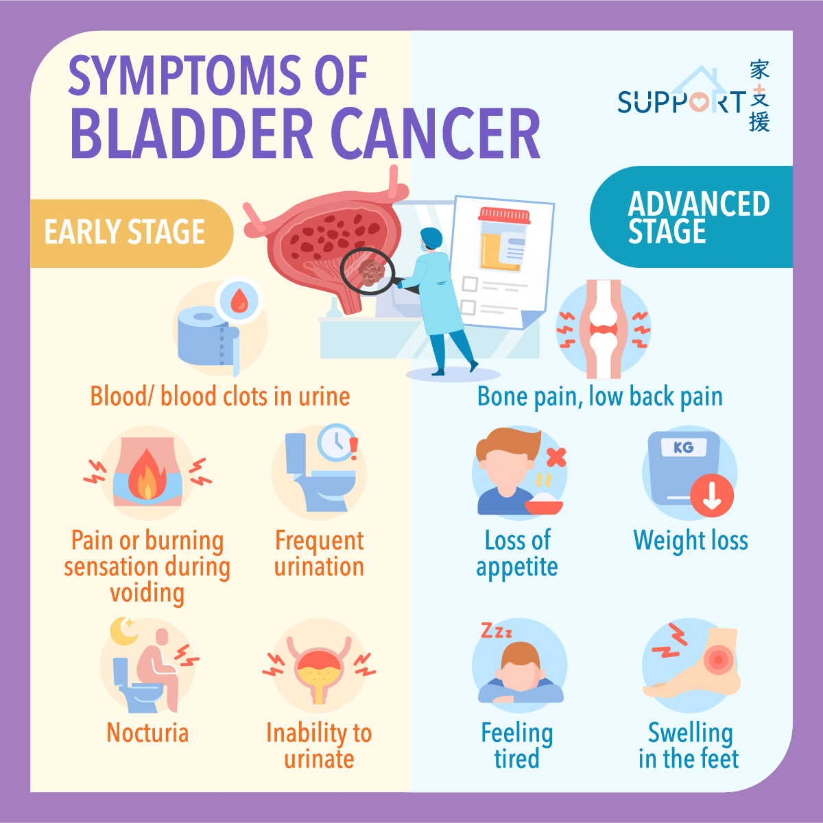
  • The most common symptom is blood in the urine (i.e. haematuria), which is usually painless.
    • You may see blood in your urine occasionally. 
    • Sometimes there are trace amounts of blood in the urine that can only be detected by a test.
  • Other common symptoms of bladder cancer include:
    • Frequent urination
    • Pain or burning sensation during urination
    • Having an urge to urinate even if your bladder isn’t full
    • Frequent nighttime urination (nocturia)
  • When the cancer has grown large or spread beyond the bladder to other parts of the body, symptoms may include: 
    • Inability to urinate
    • Lower back pain on one side of the body
    • Pain in the abdomen
    • Bone pain or tenderness
    • Unintended weight loss and loss of appetite
    • Swelling in the feet (especially if there is extensive lymph nodes involvement)
    • Feeling tired
  • It is important to check with your doctor if you have any of these symptoms. Keep in mind that urinary tract infections, bladder stones, benign prostatic hyperplasia, or other problems related to the kidney could be the cause, rather than cancer.

Diagnosis

Bladder cancer can cause symptoms like blood in the urine (haematuria) and various urinary problems that might be noticed in the early stage of the disease. If you have symptoms that suggest bladder cancer, your doctor has to find out if they are due to cancer or another condition by taking samples of your urine.

The following tests may also be performed to diagnose bladder cancer: 

  • Cystoscopy and biopsy
    • Under local anaesthesia, a cystoscope (a thin, tube-like instrument with a light and a lens for viewing) is inserted through the urethra into the bladder. 
    • Fluid (e.g. normal saline) is used to fill the bladder. Images of the inner wall of the bladder and urethra will be generated for inspection of abnormal areas.
    • Tissue samples may be removed for biopsy to diagnose bladder cancer and other conditions. 
    • After cystoscopy, you may have blood in urine, burning sensation and discomfort during voiding, or more frequent urination. These side effects usually last for a few days. If the symptoms persist or are getting worse, please contact your healthcare professionals.


Tests to stage bladder cancer 

  • Computed tomography (CT) scan of the abdominal and pelvic regions
    • Can detect tumours that have spread outside the bladder. 
    • Tumours of size < 1 cm may not be detected. 
    • Contrast medium or dye should not be used in patients with impaired kidney function.
  • Magnetic resonance imaging (MRI)
    • Detailed pictures of areas inside the body (particularly soft tissues) can be generated for assessing tumours that metastasize outside the bladder to surrounding lymph nodes. 
    • Compared with CT scan, MRI can see the soft tissue of the pelvis more clearly. This is especially important if surgery is planned.
  • Positron emission tomography (PET) scan
    • PET-CT scan can give detailed images to detect any spread outside the bladder or metastasis to other organs.
  • Bone scan
    • For detection of bone metastasis. 

Types

Urothelial carcinoma 

  • This is the most common type of bladder cancer. 
  • The cancer starts in the urothelial cells that line the inside of the bladder. 
  • Urothelial cells are also present in other parts of the urinary tract, such as the renal pelvis, ureters and urethra. Patients with bladder cancer sometimes have tumour in these areas. Therefore, the whole urinary tract should be screened for any tumours.
  • There are different subtypes of urothelial carcinoma, but these subtypes would not affect the treatment options. 

Other types:

  • Squamous cell carcinoma, accounting for 3-5% of bladder cancer cases
  • Adenocarcinoma
  • Small cell carcinoma
  • Sarcoma

Staging and grading

  • TNM staging system is commonly used for the classification of malignant tumours. 
    • Tumour (T) describes the size of the tumour, and whether the tumours have invaded the bladder lining, muscle layer, fat layer, or outside the bladder. 
    • Node (N) indicates whether the cancer cells have spread into the lymph node(s).
    • Metastasis (M) denotes whether the cancer cells have metastasized to other organs.
  • Bladder cancer can also be described as stage 0 to stage 4.
    • Stage 0 (early stage): non-invasive bladder cancer cells that are found in tissues lining the inside of the bladder but have not invaded the bladder wall.
    • Stage 1: a form of non-muscle-invasive bladder cancer that has spread to the connective tissues but has not reached the muscle layer of the bladder.
    • Stage 2: muscle-invasive bladder cancer, where the cancer has spread through the connective tissues to the muscle layers of the bladder.
    • Stage 3: cancer has grown all the way through the bladder muscles and bladder wall into the layer of fat surrounding the bladder and may have spread to the reproductive organs (prostate, seminal vesicles, uterus, or vagina) and the lymph nodes nearby, but not yet metastasized to the pelvis and abdominal wall.  
    • Stage 4: cancer has spread to the abdominal wall or pelvic wall, distal lymph nodes, and other parts of the body, such as bone, lung, or liver. 
  • Notably, if bladder cancer has invaded the muscle layer, more aggressive treatment is needed.
  • Stages 0 and 1 are cancers that have not invaded into the muscle layer while stages 2, 3, and 4 bladder cancers have already invaded into the muscle layer of the bladder wall.


Grading of bladder cancer 

  • Low-grade bladder cancer cells: tend to grow and spread more slowly than high-grade cancer cells, thus having a relatively low recurrence rate after treatment. 
  • High-grade bladder cancer: tends to grow and spread more quickly than low-grade bladder cancer. High-grade cancers usually have a worse prognosis than low-grade cancers and may need more aggressive treatments. Cancer may recur after surgery. 

Treatment for non-metastatic disease (NMIBC)

Non-muscle Invasive Bladder Cancer (NMIBC)

  • Early stage bladder cancer (stage 0 or stage 1): cancer cells have not invaded the muscle layers of the bladder. 
    • The 5-year survival rate was 96%. 
    • Nearly half of the patients were diagnosed with bladder cancer at the early stage. 
    • The 5-year survival rate reduced to 70% if the bladder cancer was invasive. 
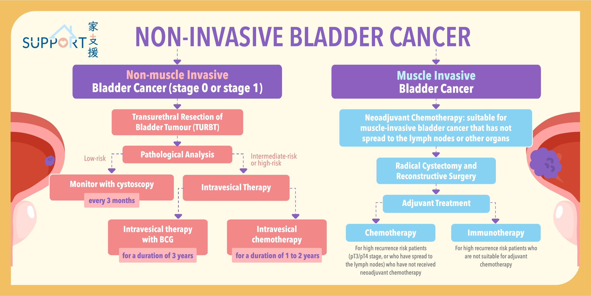
  • Early stage bladder tumours can usually be removed by Transurethral Resection of Bladder Tumour (TURBT).
    • Performed under general anaesthesia. 
    • The procedure can be completed in one hour, using heat from electricity or laser to remove the tumours. 
    • Biopsy will also be performed. Resected tissues will undergo pathological analyses. 
    • Surgical risks include blood in urine and bladder infections.
  • Intravesical chemotherapy is usually given within 24 hours of TURBT. Intravesical therapy is the instillation of medicine directly into the bladder through the urinary catheter. The aim of instillation of chemotherapy into the bladder is to clear any cancer cells that may left behind after surgery and to prevent any new cancer cells from growing.
  • Sometimes, a second, more extensive TURBT is done (usually a few weeks later) to ensure all the cancer has been removed and there is no muscle invasion.
  • If the tumour carries very high-risk features of recurrence, radical cystectomy (surgery to remove the whole bladder) may be required.
  • After TURBT, further treatment options depend on the risk of bladder cancer, such as the number, size, depth and grade of the tumours.


Low-risk NMIBC

  • After TURBT, further treatment is usually not necessary. 
  • Cystoscopy should be performed regularly to check for any recurrence. This is usually done every 3 months to start with, then less frequently with time.


Intermediate-risk or high-risk NMIBC

  • These tumours are at a higher risk of recurrence.
  • Intravesical therapy (either BCG or chemotherapy) is usually recommended after TURBT. 
  • Intravesical therapy with BCG:
    • It is usually started about 3 to 6 weeks after TURBT.
    • Intravesical BCG is given weekly for 6 weeks. Depending on the risk of recurrence, your doctor may also discuss with you on continuing the intravesical BCG with a less frequent schedule up to three years, which is called maintenance therapy.
    • Side effects: fever, chills, pain or difficulty in voiding, urge to void, blood or small particles in urine.
  • Intravesical chemotherapy:
    • Intravesical chemotherapy is the installation of chemotherapy, usually Mitomycin C, into the bladder after TURBT.
    • It may be given if intravesical BCG is not effective and its schedule is similar to intravesical BCG: once a week for 6 weeks, then monthly for 1-2 years.
    • Since the chemotherapy only goes into the bladder and not through the blood vessel to other parts of the body, it usually does not cause side effects that systemic chemotherapy may cause.

Treatment for non-metastatic disease (MIBC)

1. Surgery: Radical cystectomy

  • Radical cystectomy (RC) is a surgical procedure to remove the bladder and surrounding tissues, often used to treat muscle-invasive bladder cancer (MIBC).
  • When is radical cystectomy needed? Doctors recommend radical cystectomy if you have:
    • Advanced Bladder Cancer: This means the cancer has spread into the bladder wall (stages T2 to T4a) but hasn’t spread to lymph nodes or other parts of the body.
    • High-Risk Non-Muscle-Invasive Bladder Cancer: If other treatments haven’t worked for your cancer, you may need this surgery.
    • Research indicated that delays in undergoing radical cystectomy can negatively impact survival rates. It’s crucial for patients to proceed with surgery as soon as it’s deemed necessary to optimize their chances of recovery.


How is the Surgery Performed?

  • The procedure typically involves removing:
    • The bladder
    • Surrounding lymph nodes
    • In men, the prostate and seminal vesicles
  • There are various surgical techniques aiming at preserving sexual function. Some options include:
    • Prostate-Sparing Cystectomy: Preserves part or all of the prostate.
    • Capsule-Sparing Cystectomy: Preserves the prostate capsule while removing the bladder.
    • Seminal-Sparing Cystectomy: Preserves seminal vesicles and related structures.
    • Nerve-Sparing Cystectomy: Only the nerves are preserved to help maintain sexual function.
  • Radical cystectomy is performed under general anaesthesia, which puts you into a deep sleep. There are different ways the surgery can be done:
    • Open Surgery: This involves making a long cut in your belly to remove the bladder.
    • Robot-Assisted Surgery: This is a type of laparoscopic surgery where the surgeon uses robotic arms to perform the operation. It often results in less pain and quicker recovery because of the smaller cuts. However, studies showed that robot-assisted radical cystectomy and open radical cystectomy had a similar 90-day complication rate, surgical margin rates, oncology outcomes and quality of life outcomes.


Reconstructive Surgery After Radical Cystectomy

  • If your bladder is removed, you’ll need a new way to store and pass urine. Here are some options:
    • Incontinent Diversion: A small piece of your intestine is used to create a passage (ileal conduit) that connects the ureters to an opening (stoma) on your belly. Urine drains into a bag that you need to empty regularly.
    • Continent Diversion: A pouch is made from a piece of intestine that is connected to the ureters and has a stoma on your belly. You can empty this pouch several times a day using a thin tube (catheter). This method doesn’t require an external bag.
    • Neobladder: A new bladder is created from a piece of intestine and connected to the urethra, allowing you to urinate like before. You may need to urinate on a schedule, as you might not feel the urge.


Risks and Side Effects of Cystectomy

  • Reactions to anaesthesia
  • Bleeding
  • Blood clots in the legs (deep vein thrombosis) or lungs (pulmonary embolism)
  • Damage to nearby organs
  • Infection
  • Wound pain


2. 
Neoadjuvant Chemotherapy

  • Neoadjuvant chemotherapy, which is given before surgery, offers several important benefits for patients with muscle-invasive bladder cancer (MIBC). 
  • Here are the key advantages supported by data:
    • Improved Pathological Response Rates
      • Neoadjuvant chemotherapy can lead to significant reduction in cancer present at the time of surgery:
      • Studies showed that about 30-50% of patients who received this treatment had no remaining cancer (called ypT0) when their tissues were examined after surgery.
      • Achieving a response of ypT0 or ypT1 significantly lowered the chances of cancer recurrence. 
    • Increased Overall Survival
      • Research indicated that neoadjuvant chemotherapy can improve survival rates:
      • A review of 11 studies involving over 3,000 patients found that those who received neoadjuvant chemotherapy had an 8% better chance of surviving for five years compared to those who only had surgery.
      • In specific trials, patients treated with neoadjuvant chemotherapy experienced a 16% reduced risk of death, with 10-year survival rates improving from 30% to 36%.
    • Enhanced Surgical Outcomes
      • Studies indicated that the rate of complications after radical cystectomy was similar for those who received neoadjuvant chemotherapy and those who did not. For example, in certain trials, 86% of patients who had neoadjuvant chemotherapy were able to undergo surgery, which was almost the same as the 87% who did not have chemotherapy.
    • Better Tolerability of Treatment
      • Many patients tolerated chemotherapy better when it was given before surgery, leading to improved adherence to treatment plans.


Indications for Neoadjuvant Chemotherapy

  • Neoadjuvant chemotherapy is recommended for patients:
    • With muscle-invasive urothelial carcinoma (MIBC) that has not yet spread (cN0M0) and deemed resectable. 
    • Suitable for cisplatin-based chemotherapy.


Common Chemotherapy Regimens

  • The most common chemotherapy regimens used in this setting are cisplatin-based combinations. Some examples include:
    • Methotrexate, Vinblastine, Adriamycin, and Cisplatin (MVAC)
    • Gemcitabine and Cisplatin (GC)
    • These regimens usually consist of 3 to 6 cycles, depending on the specific treatment plan.


Side Effects of Chemotherapy

  • Nausea and vomiting
  • Loss of appetite
  • Fatigue
  • Hair loss
  • Mouth sores
  • Diarrhea
  • Constipation
  • Increased risk of infection (due to lowered white blood cell counts)
  • Anaemia (low red blood cell counts)


Neoadjuvant immunotherapy

  • There is promising evidence that adding immunotherapy to chemotherapy may enhance response rates and improve survival. 
  • Currently, three studies are ongoing to further investigate this approach. 
  • However, these treatments have not yet received approval, and we are still waiting for final results and official endorsement.


3. Adjuvant treatment

  • Adjuvant chemotherapy
    • Adjuvant chemotherapy is administered after surgery to reduce the risk of cancer recurrence. 
    • Adjuvant cisplatin-based chemotherapy may improve disease-free survival (DFS) and overall survival (OS) for high-risk patients (those with pT3, pT4, and/or lymph node positive disease) who did not receive neoadjuvant treatment. 
    • However, many trials were underpowered, making it difficult to draw definitive conclusions.
    • Moreover, challenges exist:
      • It is difficult to determine how well the tumour responds to chemotherapy, leading to potential overtreatment.
      • Additionally, some patients may experience complications after surgery that can delay or complicate chemotherapy.
      • Overall, recent studies showed a slight improvement in survival of about 6% at five years, but many trials had methodological issues. 
      • Doctors may discuss with you about adjuvant chemotherapy if your disease is more advanced (e.g. T3/4 disease and/or with pelvic lymph node involvement after surgery) and no neoadjuvant chemotherapy was given before surgery.
  • Adjuvant immunotherapy
    • Adjuvant immunotherapy uses drugs that block certain proteins in the immune system to help fight bladder cancer after surgery.
    • There are three phase III trials for patients with muscle-invasive urothelial carcinoma:
      • Nivolumab: The CheckMate 274 trial demonstrated significant improvement in disease-free survival (DFS) for patients receiving nivolumab compared to those on a placebo (20.8 months vs. 10.8 months, 6-month DFS: 74.9% vs. 60.3%). For patients with high PD-L1 expression (≥1%), the benefits were even more pronounced. Importantly, patients did not experience a decline in their quality of life compared to those on placebo.
      • Atezolizumab: In the IMvigor010 trial, patients receiving atezolizumab had a median DFS of 19.4 months, while those under observation had 16.6 months. However, this difference was not statistically significant.
      • Pembrolizumab: Results from ongoing trials evaluating its effectiveness are still pending.
  • The FDA has approved nivolumab as an adjuvant treatment for high-risk urothelial carcinoma (UC) patients post-surgery. 
  • The EMA also approved it for adults with muscle-invasive UC who have a PD-L1 expression level of 1% or higher.
  • Possible side effects of immune-checkpoint inhibitors:
    • Fatigue
    • Nausea
    • Loss of appetite
    • Fever
    • Urinary tract infections (UTIs)
    • Rash
    • Diarrhea
    • Constipation
  • Less common, but serious side effects can include:
    • Infusion reactions: Symptoms like fever, chills, rash, dizziness, and trouble breathing may occur during treatment. Report these to your healthcare team immediately.
    • Autoimmune reactions: These drugs can cause the immune system to attack healthy organs, leading to life-threatening complications in the lungs, intestines, liver, and other areas.


4. External Beam Radiotherapy

  • Radiotherapy is typically considered for patients who are not fit for cystectomy (surgery to remove the bladder) or chemoradiation. 
  • It can also be used to manage bleeding from the tumour when other treatments are not effective.

Evidence and Benefits:

  • Curative EBRT: 64–66 Gy or 55 Gy in 20 fractions.
  • Current techniques in radiotherapy offer better targeting of the bladder while minimizing exposure to surrounding healthy tissues. 
  • The recommended total dose for curative external beam radiotherapy (EBRT) is 64–66 Gy
  • An alternative is moderately hypofractionated EBRT, delivering 55 Gy in 20 fractions, which has shown similar effectiveness with less toxicity. 
  • Modern techniques have improved safety, with severe side effects occurring in fewer than 5% of patients.
  • Palliative Options: Hypofractionated regimens such as 21 Gy in 3 fractions can provide quick relief from symptoms, including haematuria (blood in urine).


Side Effects:

  • Common side effects include fatigue, urinary irritation, and acute diarrhea, which is reduced with modern techniques. 
  • Long-term complications are rare, but patients should report any new symptoms to their healthcare team.

Treatment for Metastatic Bladder Cancer

  • When choosing systemic treatment for bladder cancer, doctors consider several important factors to ensure the best approach for each patient:
    • Performance status: Patients are assessed based on their performance status, which measures how well they can carry out daily activities. This is often scored from 0 (fully active) to 4 (completely disabled). A score of 0-2 usually indicates that a patient can tolerate more aggressive treatments.
    • Kidney Function: A Glomerular Filtration Rate (GFR) of 30 mL/min or higher is often required for platinum-based chemotherapy.
    • Comorbid Conditions: Other health issues, such as heart problems or significant hearing loss, can influence treatment options, guiding the choice between cisplatin, carboplatin, or immunotherapy.


First-line systemic treatment options:

1. Enfortumab Vedotin Plus Pembrolizumab

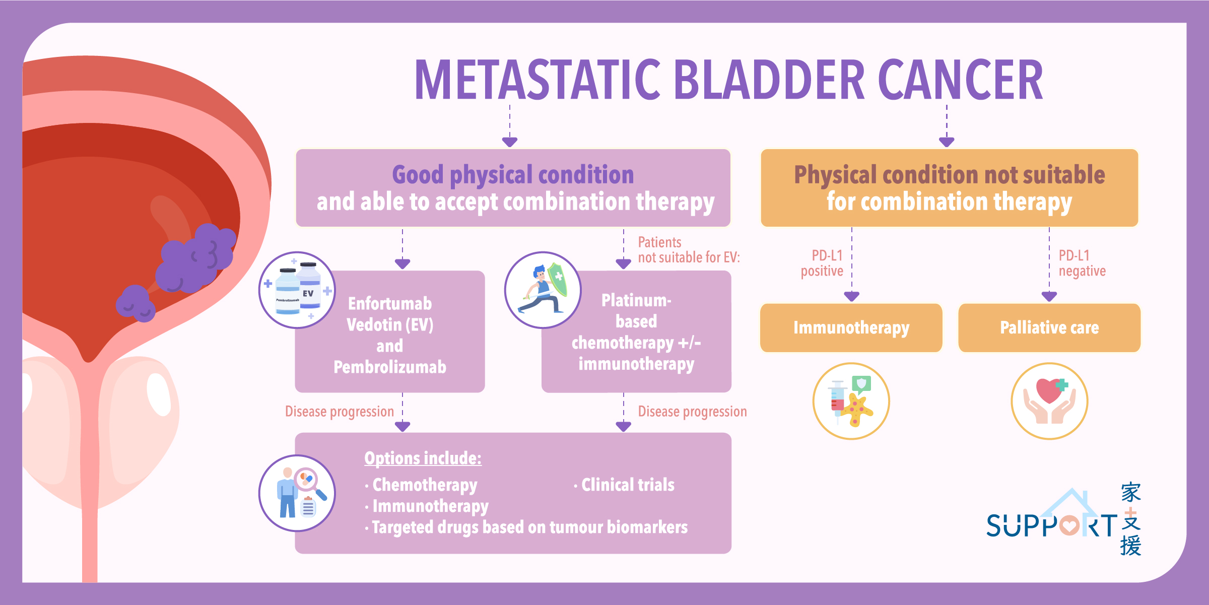
  • The combination of enfortumab vedotin (EV) and pembrolizumab is now the standard treatment for patients eligible for combination therapies. 
  • This approach is based on the EV-302/KEYNOTE 39A trial, which showed significant improvements in progression-free survival (PFS) and overall survival (OS) compared to traditional platinum-based chemotherapy. Patients receiving EV plus pembrolizumab had a median PFS of 12.5 months and median OS of 31.5 months, with a response rate of 67.7%, including 29.1% achieving complete remission.
  • Administration:
    • Enfortumab vedotin: an antibody–drug conjugate directed against nectin-4
      • Given intravenously
      • Given every 3 weeks, on Day 1 and 8
      • Potential side effects: Skin reactions, high blood glucose, inflammation of the lung, peripheral neuropathy, hair loss, loss of appetite, extravasation (drug leaking out of your vein and into the surrounding tissues), eye problems, sepsis, cardiac toxicities, and urinary tract infections. 
    • Pembrolizumab: an immune checkpoint inhibitor
      • Given intravenously
      • Given every 3 weeks, on Day 1

 
2. 
Platinum-based chemotherapy +/- immunotherapy (Nivolumab)

  • Cisplatin-based chemotherapy with gemcitabine is a common treatment for metastatic bladder cancer. The median survival of platinum-based chemotherapy is around 12-14 months. Although cisplatin may have more side effects compared with carboplatin, carboplatin-containing chemotherapy is not considered equivalent to cisplatin-based regimens for patients who can tolerate cisplatin. Comparative analyses showed that carboplatin resulted in lower response rates and shorter survival.
  • If you are fit for cisplatin, your doctor may discuss with you adding nivolumab on top of cisplatin and gemcitabine. In the CheckMate 901 trial, adding nivolumab to gemcitabine and cisplatin (GC), followed by nivolumab maintenance for up to 24 months, was compared to GC alone. 
  • Key findings included:
    • Improvement in median progression-free survival (PFS): Median PFS was 7.9 months for the nivolumab group versus 7.6 months for GC alone.
    • Improvement in overall survival (OS): Median OS was 21.7 months in the nivolumab group compared to 18.9 months for GC alone.
    • Higher response rates: The response rate was higher with the combination (57.6% vs. 43.1%), and 21.7% of patients achieved complete remission, lasting an average of 37.1 months.
  • Administration of GC and Nivolumab:
    • Route: Intravenously
    • Given every 3 weeks
    • Side effects of gemcitabine and cisplatin: nausea and vomiting, fatigue, hair loss, mouth sores, diarrhea, GI upset, renal impairment, electrolyte imbalances, and low blood cell counts, which can lead to anaemia, increased risk of infections, and easy bruising or bleeding. 

 
3.
Maintenance immunotherapy

  • Maintenance therapy with immunotherapy after platinum-based chemotherapy can be beneficial. 
  • In the JAVELIN Bladder 100 study, patients with stable disease after 4-6 cycles of chemotherapy, who received avelumab had an average overall survival of 21.4 months, compared to 14.3 months with supportive care. 
  • While 29% patients experienced immune-related side effects, most reported no significant impact on their quality of life


 4. Immunotherapy alone

  • Pembrolizumab can be considered for patients with metastatic bladder cancer who cannot receive platinum-based chemotherapy and have positive PD-L1 defined with CPS >/=10. 
  • In the KEYNOTE-052 study, 370 patients with advanced or metastatic bladder cancer ineligible for cisplatin were treated with pembrolizumab. The response rate was 29% with 7% achieving completion remission.
  • Atezolizumab can also be used in the same patient population with positive PD-L1 status, defined as 5% or more of immune cells in the tumour. 
  • In a separate phase II trial involving 119 patients, atezolizumab showed an overall response rate of 23%, with 9% of patients achieving complete remission.


Second-line systemic treatment:

  • The second-line systemic treatment options depend on the previous systemic treatment used and patients’ general condition. 
  • Options include:
    1. Combination chemotherapy, e.g. paclitaxel and gemcitabine 
    2. Immunotherapy if not used before, e.g. pembrolizumab, nivolumab, atezolizumab, avelumab, and durvalumab
    3. Antibody drug conjugates, e.g. Enfortumab vedotin, Sacituzumab govetican

Follow-up Care for Bladder Cancer Patients

Bladder cancer is a long-term disease that affects patients’ daily life. Patients should pay attention to the following for disease control and monitoring:

  • Regular follow-up
    • Cystoscopy assessment and related tests are typically performed every 3 to 6 months for the first two years.  
    • Subsequent follow-up monitoring may be scheduled annually. 
    • Initiate timely treatment to inhibit cancer growth once cancer recurrence is noted.  
  • Doctor visits and tests 
    • Symptoms identification (e.g. unintentional weight loss, loss of appetite, pain during urination, or haematuria) 
    • Physical examination of the abdominal region, wound, or stoma of urinary diversion for signs of infection or bleeding. 
    • Further assessments including cystoscopy, urine or blood tests, and abdominal x-ray imaging may be performed.  
  • Learn to manage urinary diversion. 
  • Talk to family members for emotional support.
  • Join patient sharing sessions to understand that they are not alone. 
  • Seek advice from healthcare professionals. 
    • Given that bladder cancer may recur, patients should be aware of any signs and symptoms and report to the healthcare team immediately.
    • Pay special attention to the following symptoms: 
      • Blood in urine, pain during urination, and frequent urination. 
  • Maintain healthy lifestyle and balanced diet
    • Eat more vegetables, drink more water, and exercise more. 

Prevention

  • There are many risk factors for bladder cancer. 
  • Some risk factors are modifiable, like smoking, lifestyle, and workplace exposures. 
  • Some risk factors cannot be modified, like age, sex, genetics and family history, race and ethnicity. 

The following strategies may reduce the risk of developing bladder cancer: 

  • Quit smoking 
    • Tobacco smoke contains cancer-causing chemicals that enter the bloodstream from the lungs and accumulate in the bladder. 
    • People who smoke are at least 3 times as likely to get bladder cancer as non-smokers. 
    • Smoking causes approximately half of all bladder cancers.
  • Avoid exposure to certain industrial chemicals such as paints, printing dye, hair dyes, and organic chemicals involved in the manufacturing of rubber and leather. 
  • Avoid exposure to diesel fumes.
  • Drink more water: 
    • People who drink plenty of fluids each day tend to have lower rates of bladder cancer. 
    • This might be because they empty their bladders more often, which prevents chemicals from lingering in their bladder.
  • Regular monitoring of arsenic level in drinking water by the government. 
    • Arsenic in drinking water has been linked to a higher risk of bladder cancer in some parts of the world. 
    • According to testing results from the Environmental Protection Department, the public water system in Hong Kong meets the standards for low and safe arsenic content.