Colorectal Cancer

Colorectal Cancer
Background 
Risk factors
Symptoms
Diagnosis
Types 
Stages
Treatment
Treatment for non-metastatic disease
Treatment for metastatic disease
Prevention
Video
References
Share

Background 

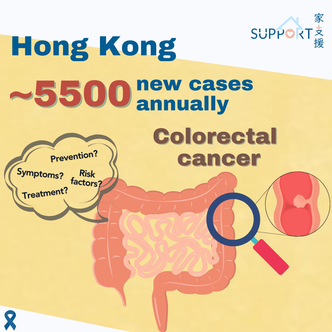
Colorectal cancer has ranked third among the ten most common cancers in Hong Kong. In 2022, there were 5,190 new cases, accounting for 14.7% of all new cancer cases. The crude annual incidence rate of colorectal cancer per 100,000 population was 70.6. Over the past ten years (2012-2022), its incidence had increased by 13.7%. The male-female ratio of colorectal cancer patients was  1.29:1. The median age at diagnosis was 69 years old and 64.3% were aged 65 or above.

Colorectal cancer was the second leading cause of cancer deaths in Hong Kong. In 2022,  2,270 deaths were caused by colorectal cancer, accounting for 15.4% of all cancer deaths.


What is colorectal cancer? 

  • The large intestine is part of the body's gastrointestinal (GI) tract or digestive system, while the colon and rectum play an important role in absorbing water and nutrition, and processing waste.
  • When there is abnormal cell growth on the bowel wall, it will emerge as polyps, ulcers or other forms of mass. Most of these are benign, but a small portion may develop into malignant tumours, leading to colorectal cancer. Cancers occurred in the colon and rectum are grouped as colorectal cancer as they are made of the same tissues.

Risk factors

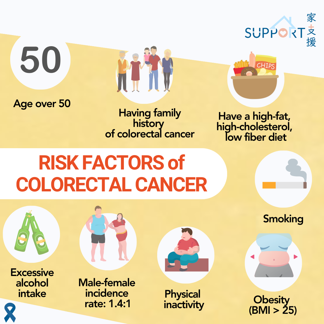
The exact cause of colorectal cancer is still unknown. People with the following characteristics are found to have a higher chance to develop colorectal cancer:

  • Age over 50
  • Having family history of colorectal cancer 
    • Colorectal cancer may be hereditary, especially from first-degree relatives
    • If many other family members such as grandparents, aunts and uncles, nieces and nephews have colorectal cancer, it is of great risk for people under the age of 60 as well
  • Suffered from or had family history of chronic colitis or colorectal polyps
  • Maintain a high-fat, high-cholesterol, low fiber diet 
    • Frequent consumption of red meat and processed meats has been consistently linked to a higher risk 
    • When a chemical in red meat called haeme is broken down in the gut, N-nitroso chemicals are formed, and these have been found to damage the cells that line the colon and rectum over time
    • These high-energy and high-fat food are also associated with a higher risk:
      • Hot or cold sugar-sweetened beverages, including soda and fruit-flavored drinks
      • Full-fat dairy products, such as whole milk cheese
      • High-fat meats, including fried chicken with skin, duck, hamburgers
  • Obesity (body mass index over 25)
  • Excessive intake of alcohol
  • Smoking
  • Physical inactivity
  • Gender: Men are more vulnerable than women, with a male-female incidence rate of 1.3:1
  • A prior history of other cancer types, e.g.
    • Previous colorectal cancer
    • Uterine cancer
    • Ovarian cancer
    • Prostate cancer 

Symptoms

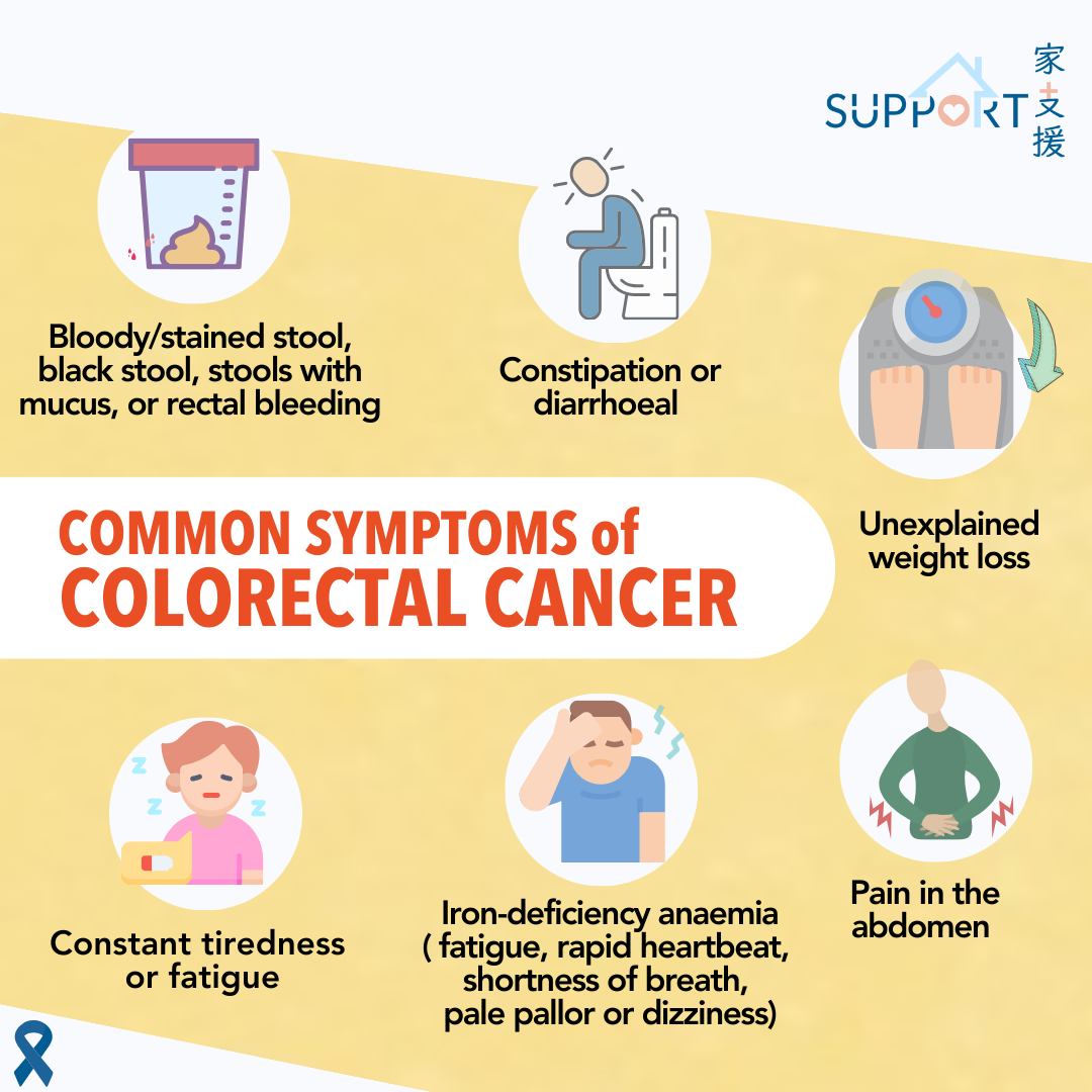
Commonly observed signs and symptoms for colorectal cancer include:

  • bloody/stained stool, black stool, stools with mucus, or rectal bleeding
  • change in bowel habits (constipation or diarrhoea), change shape of the stool (thin strip)
  • unexplained weight loss
  • pain in the abdomen (distended abdomen or colicky abdominal pain)
  • discomfort in the abdomen, including frequent gas pains, bloating, fullness or cramps
  • feeling of unfinished bowel movement
  • unexplained iron-deficiency anaemia: physical symptoms include cold hands and feet, fatigue, rapid heartbeat, shortness of breath, pale pallor or dizziness
  • constant tiredness or fatigue

Diagnosis

Colonoscopy and biopsy

  • Colonoscopy is currently the best method to examine the lower digestive tract. By means of a flexible video-endoscope, the entire length of the rectum and colon as well as the terminal portion of the small bowel could be examined. The procedure generally lasts for 10 to 45 minutes.
  • Colonoscopy inspects the entire colon. Sigmoidoscopy is used to examine the last part of colon and rectum.
  • Colonoscopy is not only useful for diagnosis. With the use of different accessory equipment, it can perform biopsy and deliver targeted procedures such as removal of polyps.
  • The specimen extracted by biopsy will be sent to pathology to determine whether the tumour is benign or malignant and help classify the tumour for guiding specific treatment options.
  • Risk and Complications
    • Minor discomforts including abdominal pain and distension are common. Major complications, including perforation, bleeding, heart and lung complications, infection or acute intestinal obstruction may happen. In general, the risk of major complications is less than 1%.  Patients should seek immediate help if abdominal pain or bloody stools appear after the procedure.


Blood tests

  • Complete Blood Count (CBC)
    • Colorectal cancer patients may become anaemic (red blood cell insufficiency) due to bleeding in the gastrointestinal tract over time.
    • CBC is to check if there is anaemia.
  • Blood chemistry tests
    • Liver function test: to check the state and condition of the liver
  • Carcinoembryonic antigen (CEA)
    • CEA is a tumour marker for colorectal cancer.
    • However, not all colorectal cancer patients will have raised CEA. CEA levels are high for only about 60% of patients diagnosed with colorectal cancer with metastasis to other organs.
    • CEA is also useful for monitoring the response of anti-cancer treatments like chemotherapy in patients with metastatic colorectal cancer.

Types 

  • Most colorectal cancers are adenocarcinomas
    • Adenocarcinoma starts in cells that make mucus along the inner lining of the colon or rectum called the mucosa. Mucus helps stool move through the colon and rectum.
  • Rarer types include:
    • Neuroendocrine tumours
    • Gastrointestinal stromal tumours (GISTs)
    • Small cell carcinomas
    • Lymphomas
    • Soft tissue sarcoma

Stages

Stage I: The cancer is contained within the muscle of the bowel wall

Stage II: The cancer has spread through the muscle of the bowel wall, but nearby lymph nodes are not affected

Stage III: The cancer has spread to nearby lymph nodes 

Stage IV: Body parts outside the bowel are affected, most often the liver or the lungs


Investigations for staging

MRI

  • MRI uses strong magnetic fields and radio waves to make cross-sectional images of the body. 
  • Endorectal MRI:
    • This is a specific type of MRI for patients with rectal cancer. The doctor will place a special probe called endorectal coil inside the rectum. The process lasts for 30 to 45 minutes and may cause discomfort.
    • This MRI can determine if the tumour has spread into nearby structures. This can help plan surgery and decide if neoadjuvant radiotherapy is needed.

CT/ PET-CT

  • Both CT and PET-CT can show detailed cross-sectional images of one’s body.
  • They are used to check if colorectal cancer has spread to other organs, such as the lymph nodes, lung or liver.

Ultrasound

  • An ultrasound uses high-frequency sound waves and their echoes to make images of different parts of the body.
    • Abdominal ultrasound: The ultrasound probe is placed over the abdomen to look for any tumours in the liver, gallbladder, pancreas, or elsewhere in the abdomen. However, it cannot be used to assess tumours in the colon or rectum.
    • Endorectal ultrasound: This test uses a special ultrasound probe to be inserted into the rectum. It is used to examine the stage of rectal cancer, including how deep the tumour has grown into the wall of the rectum and to measure how far a rectal tumour is from the anus.


Tumour biomarker testing

Microsatellite instability (MSI)

  • Normally, mismatch repair (MMR) genes correct any mistakes in DNAs that happened during cell division. When the MMR genes do not work properly, MSI can happen.
  • Tests are done to check if the cancer cells have changes in any of the mismatch repair (MMR) genes (MLH1, MSH2, MSH6, and PMS2). EPCAM, another gene related to MSH2, is also routinely checked with the 4 MMR genes.
  • Reasons for testing:
    1. To identify if the patient has Lynch Syndrome.
      • Lynch syndrome, also known as hereditary non-polyposis colorectal cancer (HNPCC), is the most common cause of hereditary colorectal (colon) cancer. People with Lynch syndrome are more likely to get colorectal cancer and other cancers, and at a younger age (before 50), including uterine, stomach, liver, kidney, brain and certain types of skin cancers.
    2. To stratify stage II patients if adjuvant chemotherapy should be recommended.
    3. To determine if immunotherapy is a possible treatment option in patients with stage IV cancers.

Gene tests

  • If cancer has spread (metastasized), doctors will check for specific gene cells which can guide on specific targeted agents for colorectal cancer.
  • Examples of gene tests to be done are KRAS, NRAS and BRAF
  • For example, for patients having colorectal cancer with KRAS mutation, they typically do not benefit from treatments with certain targeted therapy drugs, e.g. cetuximab, panitumumab
  • Some colorectal cancers that do not have mutations in the KRAS, NRAS, or BRAF genes (i.e. KRAS, NRAS, BRAF wild-type) might also be tested for HER2 or NTRK mutations.

Treatment

Treatment options and recommendations depend on several factors, including type, stage and location of colorectal cancer, the patient’s overall health condition and any other medical diseases and their preferences. Usually, a multidisciplinary team including colorectal surgeons, oncologists, radiologists and pathologists will review the patient’s general condition, type of cancer, stage of the disease and imaging results to decide on the treatment plan.

Treatment for non-metastatic disease

Stage I to III diseases are generally regarded as non-metastatic and the treatment is curative in intent. The aim of treatment is to remove all visible diseases in the body completely by surgery with or without other supplementary treatments.


Surgical Treatment

Surgery the most common treatment for colorectal cancer and represents the only curative modality. It is the removal of the tumour and some surrounding healthy tissues during an operation.

Conventional surgery: an incision is made on the abdominal wall under anaesthesia so that the operation is done under direct vision.

Laparoscopic/ robotic surgery: several viewing scopes are passed into the abdomen while a patient is under anaesthesia. The incisions are smaller and the recovery time is often shorter than with conventional surgery.

Figure: laparoscopic surgery

Colostomy: stoma, a surgical opening, is connected to the abdominal surface to provide a pathway for stool to exit the body. The colostomy can be temporary to allow the bowel wound to heal, and it can be permanent when the anal sphincter has to be removed due to cancer involvement. The stoma can be connected to a pouch to collect the faeces. 

Figure: Stoma bag

Side effects of surgery: In general, there will be pain and swelling at the site of operation. Bowel habits may also change with diarrhoea or constipation. More severe but uncommon side effects include infection and breakdown of the bowel wound. 


Perioperative therapies

Radiation Therapy: Radiation therapy is the use of high-energy x-rays to destroy cancer cells. In non-metastatic disease, it is commonly used in the treatment of rectal cancer before surgery to facilitate complete resection, improve disease control and/or preserve continence. It can be given in short-course for 5 fractions or long course in 25-30 fractions combined with chemotherapy.

Chemoradiotherapy: chemotherapy and radiation therapy can be applied simultaneously before or after surgery. It is usually given in 25-30 daily sessions for rectal cancer that has spread through the muscle wall or has involved surrounding lymph nodes. Studies have demonstrated better efficacy with fewer side effects when chemoradiotherapy is given before surgery. 

Figure: Radiotherapy planning for rectal cancer with CT scan

Side effects of radiation therapy: common acute side effects include fatigue, skin reactions, diarrhoea. Uncommon long term side effects include bowel obstruction, bleeding. Sexual problem and infertility can also occur after radiation therapy to the pelvis.

Chemotherapy: drugs that can disintegrate cancer cells by different mechanisms. It can be given intravenously via a tube placed into a vein or orally as a tablet or capsule. A patient may receive 1 drug at a time or a combination of drugs in a certain number of cycles over time. It can be combined with radiation therapy in rectal cancer to improve disease control and facilitate surgery. 

Adjuvant chemotherapy refers to chemotherapy given after surgery. Studies have proven its value in stage III disease but data is still evolving in stage II disease. In general, single-agent chemotherapy (5-FU or capecitabine) can be considered in selected patients with stage II disease while combination regimens are offered universally to patients with stage III disease who are fit to receive chemotherapy. Stage II patients who are at high risk (e.g. T4 disease, tumour perforation, <12 lymph nodes resected) for systemic recurrence may be given adjuvant chemotherapy combination. The usual duration of adjuvant therapy is 6 months but recent data has suggested 3 months of therapy will produce similar efficacy but fewer toxicities in Stage III patients with lower risk of recurrence (e.g. N1 disease)  .

Chemotherapy used in perioperative setting: 

  • 5-fluorouracil (5-FU)
  • Capecitabine
  • Oxaliplatin

Combination regimens: 

  • XELOX/ CAPOX: capecitabine and oxaliplatin
  • FOLFOX: 5-FU and oxaliplatin
  • Modified FOLFIRINOX: 5-FU, oxaliplatin, irinotecan 

In patients with advanced disease, e.g. clinically T3/4 or N2/3 disease, neoadjuvant, triplet chemotherapy using 5-FU, oxaliplatin and irinotecan may be considered. Side effects of chemotherapy: Common side effects include fatigue, nausea, vomiting, diarrhoea, mouth sores, neuropathy, skin reaction over palms and soles and increased risk of infection. Hair loss is uncommon for the above regimens in the adjuvant setting.

Treatment for metastatic disease

Metastatic disease refers to cancer cells that have already spread from the bowel to distant sites, being most common to the liver. It is defined as stage IV disease by the conventional cancer staging system. In contrast to non-metastatic disease, treatment is mostly palliative in intent. In addition to systemic treatment options, surgery and radiotherapy can be given to patients with local symptoms due to cancer involvement such as pain, bleeding and obstruction. Although the disease cannot be totally eradicated, advances in treatment have brought years of disease control and survival in the majority of patients.

In recent years, for those patients with a limited number of metastases in the liver or lungs that are amenable for surgical removal, treatment can be potentially curative. If the metastases cannot be surgically removed due to large size or quantity, chemotherapy may be given before surgery to aid resectability. Chemotherapy may be given again after surgery as well. Moreover, in some cases, if the metastasis is not able to be surgically removed, radiotherapy, ablation or embolization may be an option. 


Chemotherapy

Chemotherapy is usually given as the first line treatment if the cancer cannot be removed by surgery. It is usually the backbone of the treatment and is given in cycles over a period of time. Patients are monitored closely for toxicities and treatment response. 

Chemotherapy used in metastatic setting:

  • 5-fluorouracil (5-FU)
  • Capecitabine
  • Oxaliplatin
  • Irinotecan
  • Trifluridine and tipiracil

Combination regimens:  

  • XELOX/ CAPOX: capecitabine and oxaliplatin
  • FOLFOX: 5-FU and oxaliplatin
  • XELIRI/ CAPIRI: capecitabine and irinotecan
  • FOLFIRI: 5-FU and irinotecan

Combined with targeted therapy as first-line treatment: 

  • Epidermal growth factor receptor (EGFR) inhibitors: 
    • Target the EGFR pathway and result in stopping or slowing the growth of cancer cells. 
    • Only for patients whose tumours on the left side of the colon and have no mutation in the KRAS, NRAS and BRAF genes would benefit from this therapy. 
    • Options of combination with chemotherapy then maintenance treatment: 
      • Cetuximab
      • Panitumumab 
    • Common side effects include acneiform skin rash, diarrhoea and electrolyte disturbance.  
  • Anti-angiogenesis therapy: 
    • Targets at stopping the formation of new vessels so that cancer cells will be deprived of nutrients for growth. 
    • Given via intravenous route, in combination with chemotherapy:
      • Bevacizumab
      • Aflibercept
      • Ramucirumab
    • Patients with underlying cardiac disease or recent surgery may not be suitable for this therapy. Common side effects include minor bleeding, hypertension and protein loss in urine. Uncommon but severe side effects include perforation of bowel, thromboembolic events and wound healing problems.  


Targeted therapy

If the cancer progresses after first-line treatment, the subsequent treatment you are going to receive depends on the systemic therapy you’ve had and whether the tumour has any specific biomarkers. Examples of regimens targeting specific biomarkers are in the following figure: 


Biomarker-based treatment for colorectal cancer 

Biomarker Regimen Adminstration Side effects
BRAF V600E mutation

Encorafenib + Cetuximab

  • Encorafenib: oral
  • Cetuximab: intravenous
skin thickening, diarrhoea, skin rash, loss of appetite, abdominal pain, joint pain, fatigue, and nausea
KRAS G12C mutation
  • Sotorasib + Cetuximab or Panitumumab
  • Adagrasib + Cetuximab or Panitumumab


Sotorasib/ Adagrasib: oral nausea, vomiting, diarrhea, muscle and joint pain, fatigue, decreased appetite, and changes in liver and kidney function 
HER2 amplification
  • Trastuzumab + Pertuzumab,  Lapatinib or Tucatinib (RAS wild type)
  • Trastuzumab deruxtecan (T-DXD)
  • Lapatinib/ Tucatinib: oral
  • Trastuzumab/ Pertuzumab/ T-DXD: intravenous
  • Heart problem with lowering of left ventricular ejection fraction, diarrhea

  • Lapatinib: hand-foot syndrome and liver impairment

  • T-DXD: may cause severe lung inflammation 

NTRK gene fusion
  • Entretinib
  • Larotrectinib
  • Repotrectinib
oral
  • Dizziness, fatigue, nausea, vomiting, constipation, weight gain, and diarrhea
  • Less common but serious side effects: abnormal liver tests, increased risk for fractures, heart problems, vision changes, and confusion
RET gene fusion Selprecatinib oral
  • Decrease in white blood cell count and calcium, changes in liver function tests, high blood pressure, fatigue, changes in kidney function, and increased cholesterol
  • Less common but serious side effects: arrhythmia (QT interval prolongation), easy bleeding, difficulty in wound healing, and allergic reaction


If the cancer further progresses, options of treatment include: 

  • Fruquintinib
    • Administration: Oral
    • Each cycle : 5mg daily for 21 days of 28-day cycle
    • Side effects: Hypertension, proteinuria, bleeding tendency, liver impairment, hand-foot syndrome
  • Trifluridine and tipiracil
    • Administration: Oral
    • Each cycle: Twice a day on day 1-5 and day 8-12 of each 28-day cycle
    • Can be given together with bevacizumab to have a higher efficacy
    • Side effects: low blood count, diarrhea, fatigue, loss of appetite, nausea, abdominal pain
  • Regorafenib
    • Administration: Oral
    • Each cycle: once a day for 21 days of each 28-day cycle 


Immunotherapy

Data support its efficacy in patients colorectal cancers with MMR/MSI-H or POLE/POLD1 mutation. Options given via intravenous route include: 

  • Pembrolizumab
  • Nivolumab + ipilimumab
  • Dostarlimab 

Other modalities of treatments

In selected patients with limited metastatic involvement that are amenable to local therapies, treatment can be potentially curative, and the following specific techniques can be employed: 

  • Radiofrequency ablation (RFA):  involves the use of high-energy radio waves to heat the tumour and destroy cancer cells which have spread to the liver with a probe guided by CT scan or ultrasound. This can be used as an alternative to resection. 
    • Possible side effects include abdominal pain, infection in the liver, fever, bleeding into the chest cavity or abdomen, abnormal liver function test results.  
  • Embolisation: is used to treat liver metastases that are often too big to be treated with ablation. In an embolisation procedure, a substance is injected directly into an artery in the liver to selectively block or reduce blood flow to the tumour while leaving most of the healthy liver cells unharmed 
    • Embolisation is not suitable for patients with liver damage from diseases like hepatitis or cirrhosis. Possible side effects include abdominal pain, infection in the liver, fever, gallbladder inflammation, blood clots in the main blood vessels of the liver, abnormal liver function test results.  
    • Options:  
      • Trans-arterial embolisation (TAE) 
      • Trans-arterial chemoembolisation (TACE) 
      • Radioembolisation (ytrrium-90 microspheres) 
  • Stereotactic radiation therapy: a type of radiation therapy technique with meticulous immobilization or breathing control methods that allows delivery of a large and precise radiation dose to a small area while sparing as much normal tissue as possible. It is commonly given to cancer sites at the bones, liver, lung and brain.
  • Hyperthermic intraperitoneal chemotherapy (HIPEC): it is a technique that delivers chemotherapy directly into the abdominal space during operation. The chemotherapy is heated to enhance cancer cell kill and the procedure is most effective after surgical removal of all visible tumours in the abdomen. It is only suitable for disease that is limited to the peritoneum, a tissue lining the surface of the bowel. The data regarding its efficacy is still controversial and its practice is limited to specialized centers.

Prevention


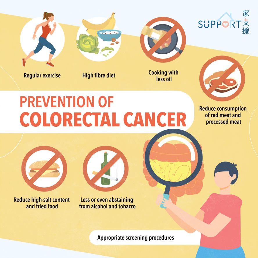
Effective ways to prevent bowel cancers include:

  • Including more dietary fibre in the diet 
    • High fibre intake promotes bowel movement, reducing constipation
    • At the same time, it prevents an overaccumulation of toxins and carcinogens (substances that cause cancer)
  • Consuming fruits and vegetables that are rich in vitamins and antioxidants
    • These vitamins and antioxidants can repair or prevent cell damage, and the fruits and vegetables themselves are rich sources of fibre 
  • Reducing consumption of red meat and processed meat
  • Cooking with less oil
    • Healthier methods include steaming, baking or grilling 
  • Reducing fried food and food with high-salt content in the diet 
  • Regular exercise to maintain a healthy weight
  • Quitting tobacco 
  • Less or even abstaining from alcohol
  • Appropriate screening procedures, see https://www.colonscreen.gov.hk/mobile/en/index.html

References

Hong Kong Cancer Registry. Overview of Hong Kong Cancer Statistics of 2022. Hong Kong Hospital Authority; Oct 2024. (accessed December 2024).

American Cancer Society. Colorectal Cancer. (accessed December 2024). 

National Comprehensive Cancer Network (NCCN) Guidelines. Colon Cancer. Version 5.2024.  

National Comprehensive Cancer Network (NCCN) Guidelines. Rectal Cancer. Version 4.2024.  

Macmillan Cancer Support. Bowel Cancer.  (accessed December 2024). 

Bowel Cancer. Cancer Council Australia (accessed December 2024). 

Special thanks to Mr. Joshua Tang, Mr. Matthew Ho-Fai Cheng (Class M23), medical student of Li Ka Shing Faculty of Medicine, the University of Hong Kong, and Dr. Ka-On Lam, Department of Clinical Oncology, the University of Hong Kong for authoring and editing this article.


Last updated on 2nd Jan, 2025.