Lung Cancer

Lung Cancer
Background
Risk factors
Symptoms
Diagnosis
Types 
Staging 
Treatment
Prevention
Clinical trials in HKU
Video
References
Share

Background

Lung cancer was the commonest cancer in Hong Kong. In 2022, there were 5,707 new cases of lung cancer in Hong Kong, accounting for 16.1% of all new cancer cases. Over the past 10 years (from 2012 to 2022), the number of new lung cancer cases rose by 23.8%. The crude annual incidence rate of lung cancer per 100,000 population was 77.7. The incidence was higher in male compared with female, with a male-to-female ratio of about 1.4 to 1. The median age at diagnosis was 70 in male and 68 in female.  

Lung cancer was also the first leading cause of cancer deaths in Hong Kong. In 2022, a total of 3,782 people died from this cancer, accounting for 25.7% of all cancer deaths.


What is lung cancer?

The lungs are a pair of organs located in the chest. Lungs consist of the bronchus, bronchioles and air sacs known as alveoli. During inhalation, lungs take oxygen into the bloodstream and deliver it to the rest of the body. During exhalation, they remove carbon dioxide from the bloodstream.

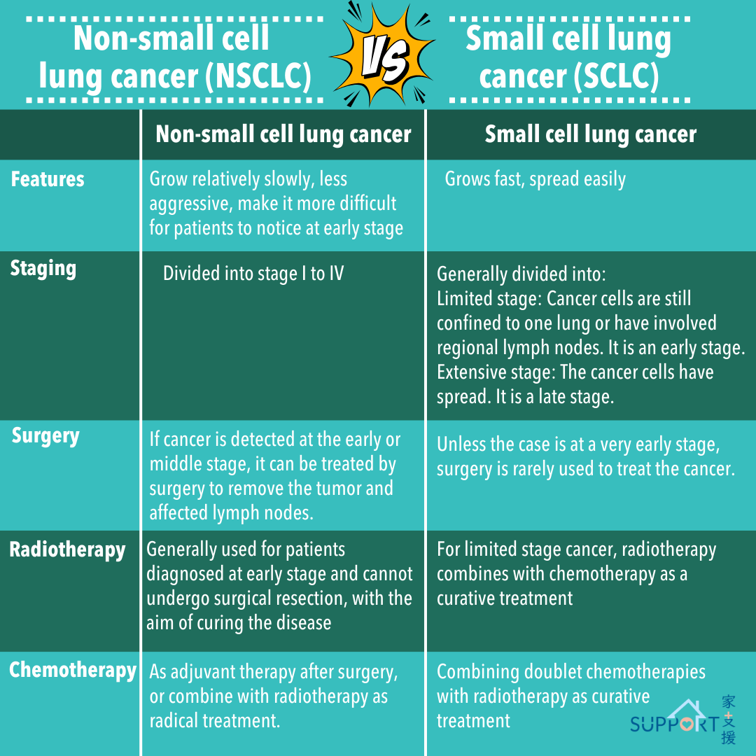
Lung cancer is a malignant tumour developed in the lower part of the respiratory system including cells in the tracheal, bronchial and bronchiolar wall.

Risk factors

The risk factors for lung cancer include:

  • Smoking 
    • In Hong Kong, 90% of male patients with lung cancer are smokers. The longer one smokes, the greater the risk for lung cancer
    • Passive smoking also increases the risk of lung cancer
    • It is estimated that 1 in 10 smokers will eventually develop lung cancer
  • Occupational exposure to certain chemicals
    • Chronic exposure to asbestos, silica and diesel exhaust
    • Handling of steel, nickel, chromium and coal gas
    • Exposure to radiation, such as radon gas in mines 
  • Air pollution
  • Age
    • People aged 50 or above are at higher risks of lung cancer
  • Family history of lung cancer (genetic predisposition)
  • Patients with weak immunity (e.g. HIV patients)

Symptoms

Symptoms of lung cancer can be divided into 3 categories: 

Respiratory symptoms

  • Persistent cough 
  • Blood-stained sputum
  • Difficulty in breathing
  • Wheezing (high-pitched, whistling breathing sounds)
  • Stridor

Symptoms caused by cancer compressing on adjacent structures

  • Chest pain
  • Hoarseness of voice
  • Difficulty in swallowing
  • Facial swelling and puffiness (as a result of superior vena cava obstruction)
  • Bone pain

Symptoms related to distant metastasis or hormonal disturbance

  • Prolonged fatigue
  • Poor appetite and unexplained weight loss 
  • Bone pain (if bone metastasis occurs)
  • Limb weakness/numbness, twitching, headache, nausea/vomiting, dizziness (if brain metastasis occurs)
  • Yellowish skin pigmentation, yellowish urine, right upper quadrant abdominal pain, abdominal distension (if liver metastasis occurs)
  • In rare cases, hormones are released from the tumour, causing low blood sodium levels or high calcium levels

Diagnosis

  • Baseline blood tests
    • Complete blood count
    • Liver function and kidney function tests
    • Electrolytes
    • Tumour markers (e.g. CEA, CA 15.3)
  • Chest X-ray
    • Usually the first test performed by doctors
    • Lung cancer tumours appear as a grey-white (radioopaquemass
    • Tumours > 1 cm may usually can be seen in a chest X-ray. However, additional tests have to be performed to confirm the diagnosis.


Investigations for histological confirmation

  • Sputum for cytology
    • A sample of sputum is examined under a microscope to look for any cancer cells.
  • Endobronchial biopsy through bronchoscopy
    • A thin, flexible tube with a camera at the end is passed through your nose or mouth down through the airways into the lung to obtain tumour samples.
    • Before the procedure, sedatives and local anaesthetic are administered.
  • Image-guided percutaneous lung biopsy
    • Usually guided by  computed tomography (CT) scan, a needle is punctured into the lungs to obtain tissue samples for microscopic examination.
    • Before the procedure, local anaesthetic may be administered.
  • Endobronchial ultrasonography-guided transbronchial needle aspiration
    • This procedure combines ultrasound and bronchoscopy to get biopsies from the mediastinal lymph nodes.
  • Thoracoscopy / Mediastinoscopy / Pleuroscopy
    • Small incision is made into the chest wall for insertion of a tube and a video camera. This allows visualisation inside the chest for lung tissue, lymph node and pleural biopsy.

Types 

Lung cancer is broadly classified into 2 main types:

A. Non-small cell lung cancer (NSCLC)

  • NSCLC accounts for more than 80% to 85% of lung cancer cases.
  • NSCLC affects the cell that line the tubes into the lungs (main bronchi) and smaller airways.
  • NSCLC can be further subclassified into 4 types 
    • Adenocarcinoma
    • Squamous cell carcinoma
    • Large cell carcinoma
    • NSCLC-NOS (not otherwise specified) e.g. adenosquamous carcinoma, sarcomatoid carcinoma

B. Small cell lung cancer (SCLC)

  • SCLC constitutes around 10% to 15% of all lung cancers
  • Tends to start in the more central part of the lungs.
  • Spreads more quickly than NSCLC.
  • Strongly linked with cigarette smoking.
  • Surgery is not often used for this type of tumour as it usually spreads early.
  • Best treated with drugs (chemotherapy), usually combined with radiotherapy.

C. Other rarer types:

  • Rarer histological subtypes include lymphoepithelioma-like carcinoma, neuroendocrine tumours, lymphoma, sarcoma etc.
    • More recently, after the discovery of the genomic aberrations, lung cancer can be classified according to its mutational subtype or expression level of the programmed death-ligand 1 (PD-L1) protein from a therapeutic point of view.
    • Common genomic aberrations found in lung cancer include EGFR, ALK, ROS1, RET, HER2, BRAF, MET, NTRK, and KRAS.

Staging 

  • Computed Tomography (CT) scan
    • Allows doctors to determine the location and size of the tumour, the extent of mediastinal lymph node involvement, and check if the tumour has spread to other parts of the body, such as the brain, thorax and abdomen.
  • Magnetic Resonance Imaging (MRI)
    • To determine if lung cancer has spread to the brain or bones.
  • Positron Emission Tomography with integrated CT scan (PET/CT)
    • Can be used to check for any spread of the cancer to other parts of the body, such as the bone, liver and pleura.
  • Bone scan
    • To show the spread of the cancer to the bones. 


A. Non-small Cell Lung Cancer (NSCLC)

  • The American Joint Committee on Cancer/Union International for Cancer Control (UICC) Staging Classification is the most commonly used staging system for lung cancer 
  • Like other solid cancers, the overall stage of NSCLC is based on the following factors:
    • Size and/or extent of invasion of the primary tumour (T)
    • Number and/or extent of spread of the lymph nodes (N)
    • Presence of spread (metastasis) to distant sites (M)

There are 4 stages of NSCLC based on the different category of T, N, and M.

Stage I

  • The cancer has not spread to the lymph nodes.
  • It is subdivided into IA and IB based on the size of the tumour. 
    • Stage IA: < 3 cm in size
    • Stage IB: 3 to 4 cm in size

Stage II

  • It is subdivided into IIA and IIB.
    • Stage IIA: tumour is 4 to 5 cm in size and has not spread into the lymph nodes nearby
    • Stage IIB: tumour is either < 5 cm in size and has spread into the lymph nodes; or 5 to 7 cm in size but has not spread into the lymph nodes

Stage III 

  • It is subdivided into IIIA, IIIB and IIIC according to the size of the tumour and the lymph nodes that the tumour has spread to.
    • Stage IIIA and IIIB: tumour may not be easily removed with surgery
    • Stage IIIC: tumour is impossible to remove with surgery 
  • The tumour has not spread to distant parts of the body.

Stage IV

  • It is subdivided into IVA and IVB.
    • Stage IVA: tumour has spread to nearby structures in the chest (e.g. the other lung, fluid surrounding the lung/heart) and/or one area outside of the chest such as to a distant lymph node or an organ such as the liver, bones, or brain
    • Stage IVB: tumour has spread to more than one part of an organ or more than one organ outside of the chest such as to distant lymph nodes and/or other organs such as the liver, bones, or brain
  • The tumour can spread to any parts of the body through the bloodstream.
  • The brain, bones, liver and adrenal glands are most likely to be affected.
  • The staging provides important information on the prognosis (survival outcome) of lung cancer.

5-year overall survival rates of stage I, II, III and IV lung cancer are roughly 75%, 60%, 40% and less than 5% respectively.


B. Small Cell Lung Cancer

A two-stage system is used to stage the disease:

  • Limited stage is when the tumour and affected lymph nodes is detected at one side of lung. The cancer is confined to an area or region that could be treated by radiotherapy with a curative intent.
  • Extensive stage is when the tumour has spread throughout two sides of the lung or other distant sites. This can only be controlled with chemotherapy and other treatment modalities with palliative intent.

Treatment

A. Treatment of non-small cell lung cancer

Stage I

  • Radical surgery aims at complete resection. Surgery offers the best chances of cure for patients with early-stage lung cancer that has not spread beyond the lungs. The curative rate of surgery stands at more than 60% among patients in the earliest stage of disease. The volume of resection depends on the status of the malignant tumour. The operation may involve the removal of a tumour with some surrounding tissue, while some may involve removal of a whole lobe or even one whole lung.
    • Depending on the size of the tumour and the presence of high-risk features, perioperative chemotherapy, immunotherapy and/or targeted therapy may be considered.  
  • Post-operative radiotherapy may be considered for incomplete resection.
  • Radical radiotherapy, such as stereotactic body radiation therapy(SBRT), can be considered for those who are not physically fit (e.g. those with significant medical comorbidities or inadequate lung function). 
  • SBRT is a technique that utilises precisely targeted radiation to the lung tumor while minimizing radiation to adjacent normal tissue.  SBRT to the lung tumour is usually given in 3-10 fractions depending on the site of the tumour (e.g. a greater number of fractions with smaller radiation dose each fraction is considered for those tumours close to important structures like heart, trachea, ribs, etc.)
  • Side effects of radiotherapy to the chest include cough, breathing difficulty, shortness of breath, hoarseness or heartburn sensation (due to inflammation of the oesophagus following radiotherapy). 

Stage II

  • Radical surgery followed by post-operative chemotherapy (usually 4 cycles of platinum-based chemotherapy).
  • For patients with tumours 4 cm or node-positive disease, preoperative (neoadjuvant) therapy an immune checkpoint inhibitor + platinum-based chemotherapy followed by one or two cycles of platinum-based chemotherapy doublet after surgery with or without continuation of singleagent immune checkpoint inhibitor may be considered. 
    • Nivolumab and pembrolizumab are immune checkpoint inhibitors used in the perioperative setting. 
  • For patients who are not candidates for immune checkpoint inhibitors due to comorbid diseases or concurrent medications, platinum-based chemotherapy doublet may be considered before surgery.
    • Examples of neoadjuvant chemotherapy include pemetrexed-cisplatin, pemetrexed-carboplatin, etoposide-cisplatin, paclitaxel-carboplatin, vinorelbine-cisplatin. 
    • Side effects of chemotherapy include nausea/ vomiting, mouth ulcers, loss of appetite, tiredness, hair loss, anemia, white cell count low, risk of infection.
  • After surgery, patients with high-risk features may be given 4 cycles of platinum-based chemotherapy doublet and the options are the same as those of neoadjuvant chemotherapy. Patients with completely resected tumours 4 cm or node-positive NSCLC may be considered for additional 1-to-3-year of systemic therapy dependent on genetic mutation status and PD-L1 expression. 
    • For patients with ALK rearrangements, oral targeted therapy using ALK inhibitor (e.g. alectinib) for up to 2 years may be considered. 
    • For patients with EGFR mutations, oral targeted therapy using EGFR inhibitor (e.g. Osimertinib) for up to 3 years can be considered.
    • An immune checkpoint inhibitor, such as atezolizumab, pembrolizumab, durvalumab or nivolumab may be given to patients with no known EGFR, ALK, RET or ROS1 mutations. One-year atezolizumab and pembrolizumab may be given to patients with PD-L1 expression 1% and have received adjuvant chemotherapy but did not receive an immune checkpoint inhibitor in the neoadjuvant setting. 
  • Post-operative radiotherapy may be considered for incomplete resection.
  • Radical radiotherapy, such as stereotactic body radiation therapy (SBRT), can be considered for those who are physically unfit for surgery (e.g. those with significant medical comorbidities or inadequate lung function). Concurrent chemoradiation may be considered for inoperable stage II patients while sequential chemotherapy/radiotherapy or radiotherapy alone may be considered for patients who are frail and unable to tolerate concurrent therapy. 

Stage III

  • For those with early and clinically occult stage IIIA lung cancer, radical surgery followed by post-operative chemotherapy (usually 4 cycles of platinum-based chemotherapy) with or without oral targeted therapy (EGFR inhibitor or ALK inhibitor) or immune checkpoint inhibitor will be considered.
  • Post-operative radiotherapy may be considered for incomplete resection.
  • Some selected patients can consider induction chemotherapy with or without immune checkpoint inhibitor or chemoradiation followed by radical surgery.
  • For patients with clinically obvious stage IIIA, IIIB, and IIIC disease; concurrent or sequential chemoradiation is usually given. The radiotherapy is delivered in 30 to 35 fractions over 6 to 7 weeks. During radiotherapy, chemotherapy is usually given in combination. Radiotherapy is typically delivered each day Monday through Friday for 15-20 minutes each time. 
  • Side effects of radiotherapy include tiredness, mild sunburn on the skin, temporary difficult and pain in swallowing, some scarring to the lungs and shortness of breath (uncommon).
  • Common chemotherapy in combination with radiotherapy:
    • Etoposide-cisplatin
    • Vinorelbine-carboplatin
    • Paclitaxel-carboplatin
  • After completion of chemoradiotherapy, immunotherapy using immune checkpoint inhibitor (e.g. durvalumab) or targeted therapy using EGFR inhibitor osimertinib can be considered as consolidation treatment. The goal is to delay the recurrence of the cancer.
  • For patients with stage IIIB/IIIC who are physically unfit to tolerate concurrent or sequential chemoradiation, targeted therapy (if actionable genomic aberrations like EGFR, ALK, ROS1 can be identified), chemotherapy, chemotherapy with immune checkpoint inhibitor, or immune checkpoint inhibitor alone (if there is high PD-L1 expression in their tumours) can be considered.


Stage IV

  • For patients with actionable genomic aberrations like EGFR, ALK, or ROS1, targeted therapy can be considered as first-line therapy.
  • Second-line therapy (after failure to first-line therapy) includes newer generations of targeted therapy, chemotherapy, or chemotherapy with immune checkpoint inhibitor, or immune checkpoint inhibitor alone.
  • For patients without actionable genomic aberrations like EGFR, ALK, or ROS1, chemotherapy, chemotherapy with immune checkpoint inhibitor or immune checkpoint inhibitor alone (if there is high PD-L1 expression in their tumours) can be considered.
  • Palliative radiotherapy can be considered for symptomatic relief (e.g. in patients with bone pain following tumour spread to the bones) 
  • Palliative care is always offered together with or without anti-cancer treatment for stage IV cancer patients.


Suggested systemic treatment for stage IV non-small cell lung cancer


Targeted therapy Examples
EGFR exon 19 deletion or exon 21 L858R mutations
  • Erlotinib (1st generation)
  • Gefitinib (1st generation)
  • Afatinib (2nd generation)
  • Dacomitinib (2nd generation)
  • Osimertinib (3rd generation)
EGFR S768I, L861Q, and/or G719X mutations
  • Erlotinib (1st generation)
  • Gefitinib (1st generation)
  • Afatinib (2nd generation)
  • Dacomitinib (2nd generation)
  • Osimertinib (3rd generation)
EGFR exon 20 insertion mutation
  • First-line: Amivantamb + platinum-based chemotherapy
  • Second-line: Amivantamab
KRAS G12C mutation 

Subsequent-line:  

  • Adagrasib
  • Sotorasib 
ALK rearrangement
  • Crizotinib (1st generation) •
  • Alectinib (2nd generation) •
  • Brigatinib (2nd generation) •
  • Ceritinib (2nd generation) •
  • Lorlatinib (3rd generation)
ROS1 rearrangement
  • Crizotinib
  • Ceritinib
  • Entrectinib
  • Repotrectinib
BRAF V600E mutation
  • Dabrafenib + Trametinib •
  • Encorafenib + Binimetinib
NTRK1/2/3 gene fusion
  • Entrectinib
  • Larotrectinib
  • Repotrectinib
MET exon 14 skipping
  • Capmatinib
  • Tepotinib
RET rearrangement
  • Selpercatinib
  • Parisetinib
HER2 mutation

Subsequent line:

  • Trastuzumab deruxtecan •
  • Trastuzumab emtansine (T-DM1)
NRG1 gene fusion 
  • Subsequent-line: Zenocutuzumab
Others
  • Bevacizumab (often used together with chemotherapy)
Immunotherapy
Depends on PD-L1 level, either use alone or concurrent with chemotherapy
  • Pembrolizumab
  • Nivolumab
  • Atezolizumab
  • Durvalumab
  • Ipilimumab
  • Cemiplimab 
  • Tremelimumab
Chemotherapy
Commonly used chemotherapy
  • Pemetrexed-carboplatin
  • Paclitaxel-carboplatin
  • Gemcitabine-carboplatin
  • Vinorelbine-carboplatin
  • Single agent chemotherapy: e.g. docetaxel


B. Treatment of small cell lung cancer

Limited stage

  • Radical surgery followed by post-operative systemic therapy with or without sequential or concurrent radiotherapy.
  • For patients with inoperable SCLC, SBRT or concurrent systemic therapy and radiotherapy may be considered.
  • Systemic therapy includes platinum-based chemotherapy doublet followed by consolidation therapy with the immune checkpoint inhibitor durvalumab.
  • In patients who responded well to initial therapy, prophylactic cranial radiation therapy to the brain in 10 daily fractions may be considered to lower the risk of brain metastases and increase overall survival.

Extensive stage

  • Primary therapy typically involves induction with an immune checkpoint inhibitor (atezolizumab or durvalumab) and platinum-based chemotherapy doublet followed by maintenance with the same immune checkpoint inhibitor.
  • Radiotherapy may be considered for selected patients e.g. brain metastases, spinal cord compression, bone metastases. 

Prevention

To prevent lung cancer, one should:

  • Quit smoking
  • Avoid exposure to second-hand smoke
  • Reduce exposure to carcinogenic substances like asbestos, silicon and radiation in workplaces and daily environment. (Refer to Ordinances such as Occupational Safety and Health Ordinance (Cap. 509) and Air Pollution Control Ordinance (Cap. 311) for more details)
  • Avoid staying in areas with serious air pollution

Clinical trials in HKU

References

American Cancer Society: Lung Cancer

Cancer Council: Types of Cancer: Lung Cancer

Hong Kong Cancer Registry. Overview of Hong Kong Cancer Statistics of 2022. Hong Kong Hospital Authority; Oct 2024

Macmillan Cancer Support: Lung Cancer

NCCN Guidelines for Patients. Early and Locally Advanced Non-Small Cell Lung Cancer, 2024.  

NCCN Guidelines for Patients. Metastatic Non-Small Cell Lung Cancer, 2024.

NCCN Guidelines for Patients. Small Cell Lung Cancer, 2024.  

NCCN Guidelines Non-Small Cell Lung Cancer Version 1.2025.  

NCCN Guidelines Small Cell Lung Cancer Version 3.2025. 

Smart patient (by Hospital Authority): Lung cancer


Special thanks to Mr. Sunny Tin-Long Wong (Class M25), Ms. Denise Hung (Class M25), medical student of Li Ka Shing Faculty of Medicine, the University of Hong Kong, Dr. Victor Ho-Fun Lee and Dr. Wendy Wing-Lok Chan, Department of Clinical Oncology, the University of Hong Kong, for authoring and editing this article.


Last updated on 4 th Jan, 2025.