Uterine Cancer

Uterine Cancer
Background
Risk factors
Symptoms
Diagnosis
Staging
Grading
Treatment
Prevention
References
Share

Background

According to the 2021 statistics data released by Hong Kong Cancer registry, uterine cancer ranked the fourth most common female cancer in Hong Kong, accounting for 6.4% of all new female cancer cases. There were 1,250 new cases of uterine cancer in 2021. On average, one out of 52 females were diagnosed with uterine cancer. The median age at diagnosis was 57 years old.

Uterine cancer was the eleventh leading cause of female cancer deaths in Hong Kong. In 2021, it claimed 128 deaths, accounting for 2.0% of all cancer deaths. 


What is uterine cancer?

The uterus is one of the female reproductive system, located in the pelvic region in the lower abdomen. The uterus is connected to left and right ovaries by 2 oviducts, and the lower part of uterus is connected to the cervix and vagina.
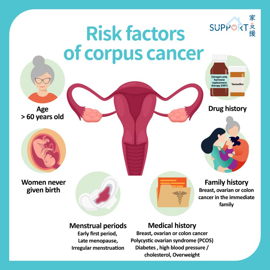
It is composed of mainly two layers: the endometrium and myometrium. The endometrium is composed of numerous capillaries (small blood vessels). Its main function is to provide an appropriate environment for the embryo implantation. Female hormones secreted by the ovaries (estrogen & progesterone) are key components towards the normal growth of endometrium. An abnormal secretion is linked with an increased risk in uterine cancer.

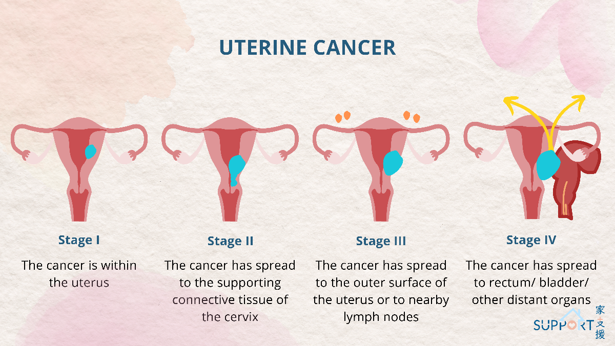
Uterine cancer is a cancer growing from the endometrium. In mid to late stage, cancerous cells will spread to structures near the uterus. It may also spread through lymph and blood to other organs like liver and bones.

Risk factors

  • Age:
    • > 60 years old, especially those after menopause
  • Menstrual periods:
    • Early first period (before 12 years old)
    • Late menopause (after 52 years old)
    • Irregular menstruation
  • Not experiencing pregnancy
  • Family history:
    • Breast, ovarian or colon cancer before 50 years old in immediate family members (mother, sister or daughter)
  • Medical history:
    • Being overweight or obese
    • Breast, ovarian or colon cancer
    • Polycystic ovarian syndrome (PCOS)
    • Endometrial hyperplasia
    • Diabetes mellitus, high triglyceride, high blood pressure and high blood glucose
  • Drug history:
    • Estrogen-only hormone replacement therapy (HRT)
    • Tamoxifen (used to treat breast cancer)

Symptoms

Patients with uterine cancer usually present with abnormal vaginal bleeding. If symptoms are discovered and medical advice is sought immediately, uterine cancer can be diagnosed at an early stage.

  • Early stage:
    • Before menopause: irregular bleeding, long and frequent period, excessive amount of bleeding
    • After menopause: sudden bleeding, abnormal discharge from vagina
  • Late stage:
    • Lower abdominal pain / distension
    • Pelvic pain during sexual intercourse or urination
    • Abnormal bowel movement (constipation)
    • Blood in vagina
    • Weight loss
    • Shortness of breath

Diagnosis

Initial diagnosis:

  • Pelvic examination
    • Finger and speculum will be used to observe any abnormalities in the vulva, vagina and cervix.
  • Endometrial biopsy
    • A thin tube will be inserted into the uterus to extract a small portion of endometrium, and the histology of the sample will be examined under a microscope.

  • Ultrasound
    • Ultrasound is useful in postmenopausal women whose endometrium should be thin.
    • If the endometrial lining is thicker than normal in postmenopausal women, further tissue evaluation is required.
    • Ultrasound is not as useful in premenopausal women, as the thickness of the endometrium varies during the ovulation / menstrual cycle.
    • Vaginal ultrasound is preferred, where a tubular probe will be inserted in the vagina.   When vaginal scan is not feasible, transabdominal ultrasound may be the alternative.

Figure: Transvaginal ultrasound
  • Hysteroscopy and biopsy / dilatation and curettage
    • Under local / general anesthetics, a fibre optic camera will be inserted into the uterus via the vagina.  Tissue sampling will be performed, and suspicious tissues will be biopsied.  The pathologists will evaluate the cells under microscope.


Further diagnosis:

  • Computer Tomography (CT) scan
    • A contrast medium will be injected intravenously, and radiographs will be taken from various angles.  Computers are used to compose a cross-sectional image, and the spread of the cancer will be evaluated.
  • Magnetic Resonance Imaging (MRI)
    • A scan of soft tissues near pelvis (lymph nodes, endometrium and myometrium) to evaluate any local spread
  • Upper abdominal ultrasound scan
    • To check for any cancer cells that might have metastasized to other organs like liver
  • Chest x-ray or CT of thorax
    • To check for any cancer cells that might have metastasized to the lungs or lining of the thoracic cavities
  • Blood-taking (tumour marker CA-125)
    • A proportion of uterine cancer cells will secrete a protein tumour marker called CA-125.
    • If it is higher than normal, it can be used to monitor treatment response and disease progression.
    • CA125 is not specific and it should only be checked after medical consultation.

Staging

According to the International Federation of Gynecology and Obstetrics (FIGO), uterine cancer can be classified into four stages:

  • Stage I
    • Cancerous cells are found within endometrium or myometrium.
    • Stage IA: Cancerous cells are within endometrium / less than half of myometrium.
    • Stage IB: Cancerous cells have spread to more than half of myometrium.
  • Stage II
    • Cancerous cells have spread from uterine to cervix (metastasis is limited to within uterus).
  • Stage III
    • Cancerous cells have spread into the pelvic cavity.
    • Stage IIIA: Cancerous cells have metastasised to the covering of the uterus (serosa), oviduct or ovaries.
    • Stage IIIB: Cancerous cells have spread to tissues outside of the uterus (parametria), or vagina.
    • Stage IIIC1: Cancerous cells have spread towards lymph nodes in pelvis.
    • Stage IIIC2: Cancerous cells have metastasised to paraaortic lymph nodes.
  • Stage IV
    • Cancerous cells have spread to colon, bladder and distal organs.
    • Stage IVA: Cancerous cells have spread to colon and bladder mucosa.
    • Stage IVB: Cancerous cells have spread to lymph nodes in the inguinal region, and distal organs; e.g. bones and lung.

Grading

Grading is a description of cancerous cell differentiation. It is used to predict the magnitude of metastasis.

Unlike normal cells, cancerous cells cannot split normally.  Therefore, pathologists can examine the tissue samples through microscopes to grade them.

  • GX: pathologists are unable to determine a grade
  • G1: cancerous cells are well-differentiated
  • G2: cancerous cells are moderately differentiated
  • G3: cancerous cells are poorly differentiated

Treatment

The current treatment options for localised uterine cancer are surgeries and radiotherapy. The two options can proceed on their own or be done together at the same time. The recovery rates of early staged uterine cancer are above 90%.


Total uterus resection

  • Preferred option for early-stage cancer
  • Procedure: resection of uterus, cervix, oviduct, ovaries, with or without part of the vagina and nearby lymph nodes. Pathological examination follows in order to determine type of cancer and any metastasis.
  • There are 4 methods of resection:
    • Abdominal hysterectomy
    • Vaginal hysterectomy
    • Laparoscopy
    • Robot-assisted
  • If the pathological reports confirm a poorly differentiated tumour, or an invasion into myometrium or cervix, then radiotherapy or chemotherapy is needed after surgery to minimise risks of cancer recurrence.
  • Risk of surgeries include anesthetic risks, post-surgical infections, bleeding, damage to bladder or intestines (risk ≤ 1-2%) and ureter, healing issues, menopausal symptoms.


Radiotherapy

  • Suitable for late-stage uterine cancer patients, and those who cannot proceed with total uterus resection
  • Radiotherapy may be considered for certain early-stage uterine cancer patients who bear risk factors for local recurrence
  • Procedure: high-energy radiation is used to eradicate cancerous cells.

1. External beam radiation therapy (EBRT)

  • A linear accelerator is used to shine radiation particles into the tumour and the pelvis to disintegrate the tumour.
  • Short term adverse effects: appear during treatment / 2-3 weeks after treatment. Possible side effects include skin reddening, fatigue, diarrhoea and pain whilst urination.
  • Medium to long term adverse effects: appear few months or even years after treatment. Possible side effects include shortening, narrowing, dryness, pain or secretion in the vagina. Other low risk but potentially serious complications include intestinal adhesions, inflammatory bowel disease, inflammation of the bladder and overactive bladder.

2. Brachytherapy (internal radiotherapy)

  • Under local / general anesthetics, a tube emitting radiation will be placed inside the vagina for treatment.
  • Several treatments may be needed. Therefore, patients may be required to be hospitalised.
  • Adverse effects:
    • Radiation may affect vagina, bladder and colon.
    • Possible side effects include shortening, narrowing, dryness, pain or secretion in the vagina. Other low risk but potentially serious complications include intestinal adhesions, inflammatory bowel disease, cystitis and overactive bladder.


Chemotherapy

  • Chemotherapy is mainly used in late-stage or recurrent uterine cancer. It helps to reduce the tumour size, slow the growth and relieve symptoms. It can also assist surgeries and radiotherapies.
  • Two common chemotherapy combinations are:
    • Carboplatin and paclitaxel 
    • Cisplatin and doxorubicin.
  • Adverse effects:
    • Seen whilst treatment but symptoms will diminish afterwards.
    • Possible side effects include fatigue, decreased immunity, nausea, decreased appetite and alopecia.
    • Doxorubicin: the heart may be affected
    • Cisplatin: the kidneys may be affected
    • Paclitaxel: the nerves may be affected; limbs may experience tingling sensations and numbness.


Others

  • Immunotherapy with or without targeted therapy might be used in selected patients.

Prevention

  • Giving birth
    • Among those who have given births, female hormones have a protective effect on the endometrium.  Therefore, risks for uterine cancer are lowered.
  • Lactation
    • During lactation, female hormones will decrease. Uterus will be less affected and results in a lower risk for uterine cancer.
    • Longer lactation period can reduce the risk of uterine cancer.
  • Regular menstruation
    • As a result of long menstrual intervals without regular shedding of the endometrium, the endometrial cells will be thickened and risks for hyperplasia and uterine cancer will be increased.
    • It is essential to use hormones, such as combined oral contraceptive pills or progestogen-only pills, to help to shed the endometrium and induce withdrawal bleeding every 2-3 months.
    • Oral progestogen or progestogen-releasing intrauterine conceptive device may also be considered for patients with heavy menstruation or endometrial hyperplasia.
  • Diet
    • Having a healthy diet can reduce risk for uterine cancer.
    • More high-fibre food should be consumed.
    • Reduce consumption of high-fat, high-sodium and high-sugar food.
    • Consume less red meat (beef, pork), manufactured food, pickled food and alcohol.
  • Prophylactic removal of uterus +/- ovaries for Lynch syndrome carriers
  • Others
    • Regular exercises
    • Weight reduction
    • Good control of diabetes and mellitus, hypertension, and hyperlipidemia

References

Hong Kong Cancer Registry, 2020, Uterine cancer

The Hong Kong Anti-Cancer Society: Uterine cancer (Chinese only)

Canadian Cancer Society: Uterine cancer

American Society of Clinical Oncology (ASCO): Uterine cancer


Special thanks to Mr. Jerome Cheuk-Him Lee (Class M25), Ms. Katrina Tung-Yee Tse (Class M25), medical student of Li Ka Shing Faculty of Medicine, the University of Hong Kong, and Dr. Ka-Yu Tse, Department of Obstetrics & Gynaecology, the University of Hong Kong for authoring and editing this article.


Last updated on 18th Dec, 2023.