Liver Cancer

Liver Cancer
Background
Risk factors
Symptoms
Diagnosis
Types 
Staging
Treatment
Treatment for localised tumour
Systemic therapy for advanced HCC
Prevention
Video
References
Share

Background

Liver cancer was the fifth commonest cancer in Hong Kong and accounted for 4.6% of all new cases in 2022. There were 1,612 new cases of liver cancer in 2022. Over the past ten years (2012-2022), its incidence had reduced by 9.9% from 1,790 in 2012.

The male to female ratio was 2.7 to 1. The crude annual incidence rate per 100,000 population was 22.  

Liver cancer was the third leading cause of cancer deaths in Hong Kong. In 2022, a total of 1,412 people died from this cancer, accounting for 19.2% of all cancer deaths.

Liver cancer is relatively difficult to cure because the cancer is usually diagnosed in the mid or late stage. Prevention is always better than cure. We should get the hepatitis B vaccination.  In addition, hepatitis B virus carriers should also attend regular follow-up to check for signs of cancer development.


What is liver cancer (hepatocellular carcinoma)?

The liver is a very important metabolic organ. It is found on the upper right of the abdomen, comprising the left lobe and the right lobe. The main functions of the liver include:

  • Producing and storing glucose for necessary use
  • Producing bile for digesting fats in food
  • Detoxifying toxins and alcohol
  • Producing proteins, blood-clotting components, antibodies and cholesterol

Liver cancer will develop when liver cells begin to mutate and split uncontrolledly. It is further divided into:

  • Hepatocellular Carcinoma (HCC), arising from liver cells 
  • Cholangiocarcinoma or bile duct cancer, arising from the cells lining the bile duct

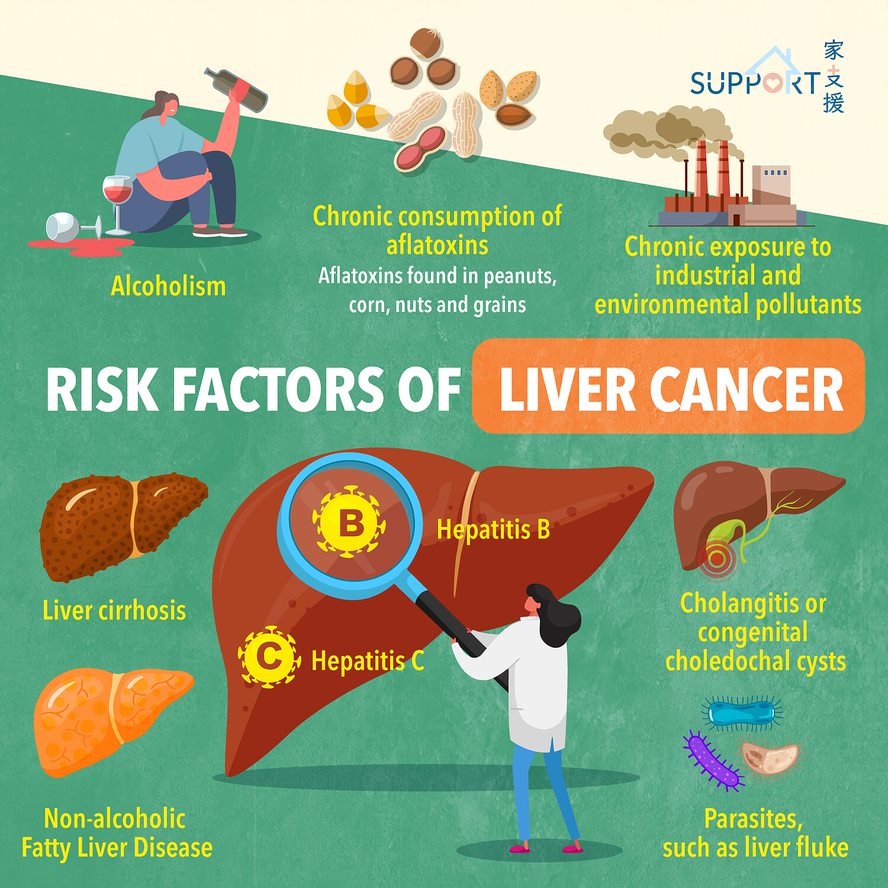
Risk factors

  • Hepatitis B 
    • The probability of liver cancer for hepatitis B virus carriers is 100 times higher than non-carriers
    • 55% of liver cancer cases around the world are caused by hepatitis B infection 
    • Hepatitis B infection is very common in Hong Kong. Around one tenth of the population are either carriers or have had a prior history of infection
      • Among these carriers, 25% of them may develop into chronic cirrhosis, which may cause liver cancer
    • It generally takes 50 to 60 years to develop into liver cancer after
      hepatitis B virus infection
  • Hepatitis C 
    • The chance of hepatitis C virus (HCV) carriers to get liver cancer is 150 times higher than non-carriers. HCV-associated liver cancer is more prevalent in western countries
  • Cirrhosis of the liver
    • Hepatitis B virus may develop into chronic hepatitis, which may further develop into cirrhosis hence become liver cancer. The actual progress of the disease varies among people and depends on how active the hepatitis virus is
    • Studies have shown that the more active the viruses are, the faster the liver cells are damaged
  • Alcoholism
    • Consuming excessive alcohol may cause liver hardening, which may then develop into liver cancer
    • The chance of alcoholic hepatitis B carriers getting liver cell cancer is 2 times higher than that of general virus carriers
  • Non-alcoholic Fatty Liver Disease (NAFLD) and Non-alcoholic Hepatosteatosis (NASH)
    • Obesity, diabetes mellitus, and other metabolic disorders may induce liver damage, causing a chain reaction leading to cirrhosis
  • Chronic consumption of aflatoxins
    • Aflatoxins found in peanuts, corn, nuts and grains 
  • Chronic exposure to industrial and environmental pollutants
    • Byproduct fumes during the manufacture of PVC plastics are known carcinogens
  • Cholangitis (inflamed bile duct) or congenital choledochal cysts
    • May lead to bile duct cancer
  • Parasites
    • In Asia, a parasite known as the liver fluke can cause chronic infections of the gallbladder and liver, leading to liver cancer

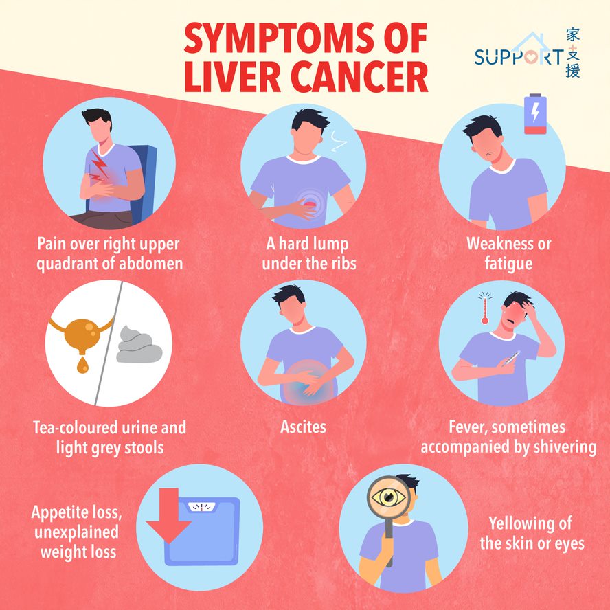
Symptoms

  • Pain, especially at the top right of the abdominal area, near the right shoulder blade, or in the back
  • Unexplained weight loss
  • Appetite loss
  • Fever, sometimes accompanied by shivering
  • A hard lump under the ribs, which could be the tumour or a sign that the liver has enlarged
  • Weakness or fatigue (lethargy)
  • Yellowing of the skin or eyes, called jaundice
  • Tea-coloured urine and light grey stool
  • Abdominal swelling caused by ascites (fluid accumulation)

Diagnosis

Blood test

  • Alpha-fetoprotein (AFP): 
    • A tumour marker for liver cancer which can be used for screening of liver cancer in high-risk groups such as patients with liver cirrhosis, chronic hepatitis B or C
    • Helps to determine prognosis, monitor response to therapy and detect recurrence
  • Liver function test:
    • Checks on liver function in order to design subsequent treatment

Ultrasound of the liver

  • Used to inspect the structure of the liver, to confirm the size and location of the tumour. It can be used to guide on the biopsy of the liver lesion. A gel is spread onto patient’s abdomen. A small device that produces sound waves is passed over the area. The sound waves are then converted into a picture by a computer. The process is painless and only takes a few minutes.

CT (computed tomography) scan of the abdomen

  • Uses a series of x-rays taken of the abdomen then fed into a computer
  • A detailed 3-D picture of the size and positions of the cancer is produced

MRI (magnetic resonance imaging) scan

  • Very similar to a CT scan, but uses strong magnetic fields instead of x-rays to build up cross-sectional pictures of the patient’s body.

Laparoscopy

  • A small, minimally invasive operation allowing the doctor to look into the liver. All conducted under general anesthesia.
  • A small cut in the lower part of the abdomen will be made to insert a laparoscope (a mini telescope)
  • Besides inspecting the liver, the laparoscope is also built to allow tissue samples (biopsy) to be taken

Liver biopsy

  • Usually done together with ultrasound to find the best site for needle insertion
  • After extracting cells from the liver tumour, the tissues will be examined under microscope to determine the presence of cancer cells

Types 

Malignant cancer of the liver can be divided into two groups: primary and secondary cancer

  • Primary cancer
    • Primary cancers are cancers that arise from the liver
    • They are further divided into hepatocellular carcinoma, cholangiocarcinoma and angiosarcoma
    • Hepatocellular carcinoma (HCC): 
      • Hepatocellular carcinoma is the most common type of liver cancer, which accounts for 75% of primary liver cancers in adults
      • The cancer arises from liver cells
      • Usually, the cancer is confined to the liver. However, there are still chances for the cancer to spread to other organs
      • Cholangiocarcinoma:
        • Cholangiocarcinoma accounts for 10%-20% of primary liver cancers in adults. 
        • The cancer arises from cells in the bile duct of the liver, a thin tube that connects the liver to the small intestine. 
      • Angiosarcoma:
        • Angiosarcoma accounts for only 1% of primary liver cancers in adults.
        • The cancer arises from blood vessels in the liver and grows quickly.
  • Secondary Cancer
    • Secondary cancers are cancers that arise in other parts of the body apart from the liver
    • Almost all cancer can spread to the liver, in which the most common ones are pancreas, stomach, bowel, breast and lung cancer

**In this chapter, we only focus on hepatocellular carcinoma (HCC).

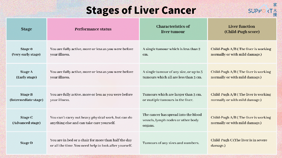
Staging

For hepatocellular carcinoma, the Barcelona Clinic Liver Cancer (BCLC) system is used to describe the stages of cancer and to recommend treatment options. The BCLC system categorises hepatocellular carcinoma based on patient’s performance status, characteristics of liver tumour and liver function (Child-Pugh score).

Treatment

Doctors will consider the following factors when designing a treatment plan:

  • Whether the cancer is a primary or secondary liver cancer
  • Age
  • General health
  • Type and size of the cancer
  • Whether it has spread beyond the liver
  • Whether the liver is affected by any other diseases, such as cirrhosis

When a tumour is found at an early stage and the patient’s liver functions normally:

  • Treatment is aimed at eliminating the cancer
  • When liver cancer is found at a later stage, or the patient’s liver is not working well, the goals may be slowing the development of the cancer and relieving symptoms to improve quality of life

Treatment for localised tumour

Disease directed treatment

Surgery, radiofrequency ablation, percutaneous ethanol injection and radiotherapy are usually recommended when the tumour is located in the liver and has not yet spread to sites outside the liver.

If the disease is in the early stage, surgery is often the first option. However, some patients may have comorbidities, or their liver functions are not fit for surgery or disease directed treatment. In this case, radiofrequency ablation or radiotherapy may be offered.


1. Surgical treatment 

  • Used to remove the tumour and surrounding affected tissues. Removal by radical surgery suits 20% of liver cancer patients whose tumours affect only one of the liver lobes and their liver functions are normal. 3-year and 5-year survival rates are 62% and 50% respectively.
  • The removal of the tumour and some surrounding healthy tissues from a portion of the liver

Likely to be the most effective, particularly for:

  • Patients with good liver function 
  • Tumours that can be safely removed from a limited portion of the liver

It may not be an option if:

  • The tumour takes up too much of the liver 
  • The liver is too damaged, the tumour has spread outside the liver, or if the patient has other serious illnesses


Two types of surgery are used to treat Hepatocellular carcinoma (HCC):

  • Hepatectomy
    • For hepatectomy, only a portion of the liver is removed. Hepatectomy can be done only if the cancer is in one part of the liver while the rest are normal
    • After hepatectomy, the remaining section(s) of the liver will take over the functions of the entire organ, and the liver may grow back into its normal size within a few weeks
    • A hepatectomy may not be possible if the patient has advanced cirrhosis, even if the tumour is small
    • Side effects of hepatectomy may include: pain, weakness, fatigue, and temporary liver failure


  • Liver transplantation 
    • Particularly effective for people with a small tumour, as both the tumour and the damaged liver are removed. However, there are few donors and patients may need to wait for a long time.
    • Liver transplant may be possible if: 
    • The patient either has a single tumour that is 5 cm or smaller, or has 3 or fewer tumours, all smaller than 3 cm
    • a suitable donor is found. 
    • After liver transplant, the patient will be closely monitored for signs of organ rejection or tumour recurrence. The patient must take medication to prevent rejection. These drugs may have side effects, such as puffiness in the face, high blood pressure, or increased body hair
    • Other serious complications of liver transplant include death from infection, organ rejection, and an increased risk of other unrelated cancer


2. Other liver directed treatment:

  • Radiofrequency ablation (RFA)
    • RFA uses electrical currents to generate heat to destroy cancer cells
    • RFA is given through skin, through laparoscopy, or open surgery while the patient is sedated
    • The technique works best for tumours less than 5cm in size.
    • Length of procedure: between 20 minutes to 2 hours, depending on the size and number of tumours
    • Side effects include mild swelling of the liver, bleeding, infection and damage on surrounding organs (bowel, stomach)


  • Percutaneous ethanol injection
    • Percutaneous ethanol injection is when alcohol is injected though the skin and into the liver tumour to destroy it. 
    • This procedure is usually done with ultrasound guidance to make sure the needle goes directly into the tumour. If the tumour grows again, the treatment can be repeated.
    • In general, the procedure is simple, safe, and particularly effective for a tumour smaller than 3 cm. However, if the alcohol escapes from the liver, a person may have brief but severe pain. So, this procedure is largely replaced by RFA.
    • Side effects include fever, bleeding, bile duct inflammation or bile duct leakage. 


  • High Intensity Focused Ultrasound (HIFU)
    • HIFU utilises a unique frequency of the ultrasound wave that can be focused on a distance from the therapeutic transducer. The accumulated energy at the focused region causes oscillation of the particles, elevating tissue temperature and inducing necrosis of the target lesion. 
    • Unlike other standard ablation methods, it does not puncture the tumour. So, it is not associated with the risk of bleeding and cancer cell dissemination.
    • HIFU works best in smaller tumours with size less than 3cm.
    • Side effects include skin reaction (usually mild), liver necrosis, liver failure and damage to surrounding tissues (e.g. bowel or stomach)


  • Stereotactic body Radiation Therapy (SBRT)
    • SBRT is to use a high precision technique to deliver a high dose of radiation to the liver tumour and keep rapid fall-off doses away from the target, thereby achieving maximum treatment efficacy with minimal toxicity to normal tissues.
    • Typically, SBRT is completed in 3-5 treatments over the course of 1-2 weeks.  This is opposed to the daily standard external beam radiation treatment that is typically given over the course of multiple weeks.  
    • Before SBRT, patient will attend a planning session and a mold will be done to immobilize the patient on the coach. A CT scan with contrast will be done for radiotherapy planning. In addition, MRI provides better tissue resolution and complementary information which are useful for target delineation. 
    • During treatment, a detector will be put on the abdomen of the patient to monitor the breathing pattern. Since the tumour may also move with breathing, it is important to gauge the breathing in order to have a tight margin around the tumour and spare surrounding normal tissues.
    • Side effects of SBRT include pain, fatigue, liver toxicity, damage to the gastrointestinal tract, biliary tract and rib fracture.


  • Selective Internal Radiation Therapy (SIRT)
    • SIRT is a special type of radiation therapy that targets liver tumours and delivers radiation from millions of tiny radioactive beads directly to the tumours.
    • The microspheres are injected through a catheter in the groin and travel through the arteries of the liver to lodge in the very small blood vessels in and around the liver tumour(s), where they emit high doses of radiation. As the microspheres only give off radiation to a small area, they target the liver tumour while causing minimum damage to surrounding healthy liver tissues.
    • Before the procedure, an arteriogram is done to check which arteries bring blood to the liver tumours. The arteriogram also tells if any arteries bring blood outside the liver or other parts of the body, such as the lungs. The radiologist will then decide whether the patient is suitable for SIRT.
    • During the procedure, a small incision is made in the patient's groin and a flexible catheter is guided into the liver through the femoral artery in the leg up to the tumour sites. The catheter is moved through the hepatic artery and positioned by the interventional radiologist to allow for targeted infusion of the SIR-Spheres microspheres to the site of the tumours. The microspheres take approximately 15 minutes to be infused, the whole procedure takes about one hour. 
    • Side effects include pain, bleeding and feve. Radiation dose to the stomach, small bowel or pancreas may cause ulceration and inflammation


  • Transarterial chemoembolization (TACE)
    • TACE is a treatment that combines two mechanisms: blocking the blood supply to the liver tumour (embolization); and delivering chemotherapy directly to the tumour. 
    • TACE is done in the x-ray department of a hospital. The procedure is usually done under local anesthesia. A thin catheter is placed into the artery in the groin (femoral artery). The catheter then goes from the femoral artery up to the hepatic artery in the liver. A dye is injected into the catheter and an x-ray (called an angiogram) is taken to find the branches of the artery that are feeding the liver tumour. The catheter is further moved up to the feeding arteries. The doctor injects a material through the catheter. The material is soaked with chemotherapy, usually doxorubicin or cisplatin. The material will then block the flow of blood to a tumour so the cancer cells die. 
    • Side effects include fever, pain, nausea and vomiting, fatigue, abnormal liver function, inflammation of the gallbladder or bile ducts, as well as liver failure especially in advanced cirrhosis.

Systemic therapy for advanced HCC

If the liver cancer has spread to other sites and is not suitable for the above localized treatment, systemic treatment can be considered for controlling the disease.

The type of medications for advanced HCC includes immunotherapy and targeted therapy.


Immunotherapy 

  • For example: Atezolizumab, nivolumab, pembrolizumab, ipilimumab, durvalumab and tremelimumab
  • The combination of Atezolizumab (a type of immunotherapy) and bevacizumab (a targeted agent that inhibit growth of blood vessels in the tumour) can be used as first-line treatment for unresectable or metastatic HCC patients.
  • The combination of Durvalumab and Tremelimumab (double immunotherapy) have been proven to be an effective first-line treatment for unresectable or metastatic HCC patients. 
  • The use of pembrolizumab, nivolumab or nivolumab-ipilimumab combination can be considered in patients who have disease progression after use of targeted agents.
  • Side effects include hypertension, fever, raised liver enzymes, proteinuria and fatigue


Targeted therapy 

  •  For example: Lenvatinib, Sorafenib, Ramucirumab, Cabozantinib, Regorafenib
  • Lenvatinib and sorafenib
    • Both lenvatinib and sorafenib are oral multikinase inhibitors. They can be used as first-line treatment for metastatic HCC.
    • Study showed that lenvatinib, when compared with sorafenib, had a better response rate and longer time-to-progression while similar overall survival.
    • Administration: oral medication, dose depends on body weight
    • Side effects of lenvatinib include high blood pressure, heart problems, deranged liver function, diarrhoea, proteinuria, hypocalcemia, changes in electrical activity of the heart (prolonged QT interval) and wound healing problems
    • Side effects of sorafenib include diarrhoea, fatigue and skin problems

Prevention

  • Limiting alcohol consumption
    • The liver is already overworked with 500 different functions, while alcohol in the blood casts more strain on the liver than normal
  • Regular work and rest periods
    • Allows the liver to function better and repair itself constantly while preventing fatty liver disease
  • Eating more fruits and vegetables for antioxidant intake
  • Safe sex with condoms and avoid sharing needles
    • 8-10% of Hong Kong’s population are carriers of the hepatitis B virus (HBV), while 0.5% carry the hepatitis C virus. 
    • Both hepatitis B and C can be transmitted through blood and reproductive fluids
  • Food storage and treatment
    • Rotten peanuts, grains and corn may produce aflatoxins. Discard as soon as signs of spoilage become apparent
    • Cook freshwater fish properly to minimise risks of liver fluke infestation
  • Screening and vaccinations
    • The hepatitis B vaccine can protect 95% of people who are vaccinated
    • Family members should be screened as well

References

Hospital Authority Hong Kong Cancer Registry, 2022

The Hong Kong Anti-Cancer Society: Liver cancer (Chinese only)

Smart Patient (by Hospital Authority): Liver cancer

American Society of Clinical Oncology (ASCO): Liver cancer

Canadian Cancer Society: Liver cancer


Special thanks to Mr. Joshua Tang, Ms. Jasmine Wing-Fong Wu (Class M23), medical student of Li Ka Shing Faculty of Medicine, the University of Hong Kong, and Dr. Chi-Leung Chiang, Department of Clinical Oncology, the University of Hong Kong for authoring and editing this article.


Last updated on 30th Nov, 2024.