Oesophageal Cancer

Oesophageal Cancer
Background
Risk Factors
Symptoms
Diagnosis
Types 
Staging 
Treatment 
Treatment for non-metastatic disease
Treatment for advanced or metastatic disease
Prevention
Clinical trials in HKU
References
Share

Background

There are about 400 new cases of oesophageal cancer in Hong Kong every year. Males are more prone to this disease than females, with the male-to-female ratio of incidence being around 4.5:1 in 2021. The incidence rate of oesophageal cancer in Asia is significantly higher than that in western countries, which is probably related to the dietary habits of Asians. The incidence rate also increases with age.

In 2021, 299 patients died from oesophageal cancer, accounting for 2.0% of all cancer deaths. The crude death rates for male and female were 7.1 and 1.5 per 100,000 population respectively.


What is oesophageal cancer?

Oesophagus

  • A long, hollow muscular tube, measuring 25-30cm, connecting the throat to the stomach
  • Part of a person’s gastrointestinal (GI) tract, also called the digestive system
  • The oesophagus does not play a role in digesting food. It pushes food and fluids from the mouth into the stomach.

Oesophageal cancer

Oesophageal cancer is a malignant tumour originated at the oesophagus. Malignant transformation of cells occurs when genetic mutation in the oesophageal tissue cells leads to uncontrolled growth and invasion of adjacent tissues. Depending on the type of cells, it is classified into squamous cell carcinoma and adenocarcinoma. In general, squamous cell carcinoma usually affects the neck (upper part) and chest (middle part) while adenocarcinoma commonly affects the junction of oesophagus and stomach (lower part).

Risk Factors

The following factors may increase risks of developing oesophageal cancer:

  • Age: Aging 60 or above causes a higher risk of developing oesphagal cancer
  • Gender: Men are 3 to 4 times more likely than women to develop oesophageal cancer
  • Smoking: Using any forms of tobacco, such as cigarettes, cigars, pipes, chewing tobacco and snuff raises the risk of oesophageal cancer, especially SCC
  • Alcohol: Heavy drinking over a long course increases the risk of squamous cell carcinoma of the oesophagus, especially when combined with tobacco use
  • Consumption very hot liquid or soups
  • Consumption of preserved or smoked food
  • Inadequate consumption of fruits and vegetables
  • Acid reflux
  • Certain rare diseases (such as achalasia) and inherited gene mutations (such as PTEN and Peutz-Jeghers syndrome (PJS))

Symptoms

The first symptom experienced by most oesophageal cancer patients is progressive difficulty in swallowing. However, patients may not be aware and may change their eating habits unconsciously. Since there is difficulty in eating, weight loss and malnutrition are often seen in patients. When one has trouble in swallowing, or experience weight loss or heartburns for unknown reasons, a consultation with the doctor should be arranged as soon as possible.

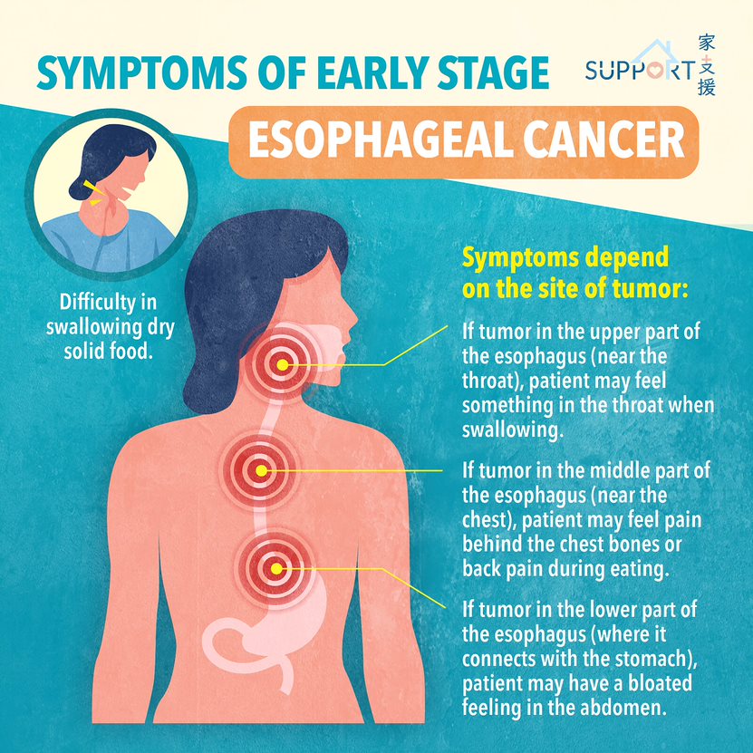
The early stage symptoms of oesophageal cancer include:

  • Difficulty in swallowing dry solid food
  • Discomfort and pain in the chest while eating
  • If tumour appears in the upper part of the oesophagus (near the throat), there will be discomfort or a foreign body sensation in the throat while swallowing food
  • If tumour appears in the middle part of the oesophagus (near the chest), pain behind the chest bones or back pain will be felt while eating
  • If tumour appears in the lower part of the oesophagus (where it connects with the stomach), the abdomen may feel bloated

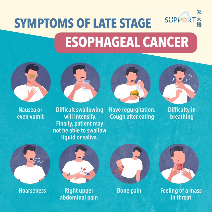
The symptoms of late stage oesophageal cancer include:

  • Intensified difficulty in swallowing. Gradually, patient will only be able to consume semi-liquid food, and patient even be unable to swallow liquid or saliva eventually.
  • Tumour in the oesophagus disturbs normal digestion in the stomach. Patients may feel nauseated, or experience vomiting and regurgitation (bringing food back up).
  • Cough after eating
  • Hoarseness
  • Other symptoms caused spread of the tumour to other parts of the body, such as a foreign body sensation in the throat, bone pain, difficulty in breathing and right upper abdominal pain.

Diagnosis

  • Upper endoscopy, also called oesophagus-gastric-duodenoscopy (OGD)
    • It is a thin, flexible tube with a light and video camera at the end, called an endoscope, which can examine the oesophagus, stomach and duodenum.
    • With the use of different types of accessory equipment, the doctor can perform biopsy and deliver surgical therapies to treat the upper gastrointestinal tract.
    • In general, the procedure lasts for 5-20 minutes, but the process may be prolonged in complex cases that require additional therapies, such as in the control of active bleeding. Patients will be carefully monitored during the procedure.
  • Endoscopic ultrasound (EUS)
    • Often done at the same time as an upper endoscopy
    • An endoscopic probe with an attached ultrasound device is inserted into the oesophagus
    • Ultrasound is used to determine if the tumour has grown into the wall of the oesophagus, the depth where the tumour is located, and whether cancer has spread to the lymph nodes or other nearby structures. 
    • An ultrasound can also be used to conduct a lymph node biopsy
  • Bronchoscopy
    • Since the oesophagus is adjacent to the airway, a bronchoscopy is needed to check if the tumour has invaded the trachea or bronchi.
    • Like the upper endoscopy, a thin, flexible tube is inserted through the nose or mouth through the trachea and bronchi.
  • Biopsy
    • Usually performed under upper endoscopy or EUS
    • During the procedure, a small sample of tumour tissue is extracted and sent for pathological review to check for any cancer cells.
    • This is the only definite way for an accurate cancer diagnosis
  • Barium swallow
    • The patient swallows a liquid containing barium followed by a series of x-rays 
    • Barium coats the surface of the oesophagus, making a tumour or other abnormalities easier to detect

Types 

  • Oesophageal squamous cell carcinoma (SCC)
    • More commonly seen in Southern China, the Middle East, South America, Western Europe and Africa
    • Usually develops in the upper and middle part of the oesophagus
    • Strongly linked with smoking and alcohol consumption
  • Adenocarcinoma
    • More commonly seen in western countries
    • Usually develops at the lower end of the oesophagus and the upper part of the stomach
    • Occurs more often in people who are in middle age, overweight and with gastric reflux
  • Rarer types of oesophageal cancer:
    • Leiomyosarcoma
    • Gastrointestinal stroma tumour (GIST)
    • Neuroendocrine tumours (NET)
    • Small cell carcinoma

Staging 

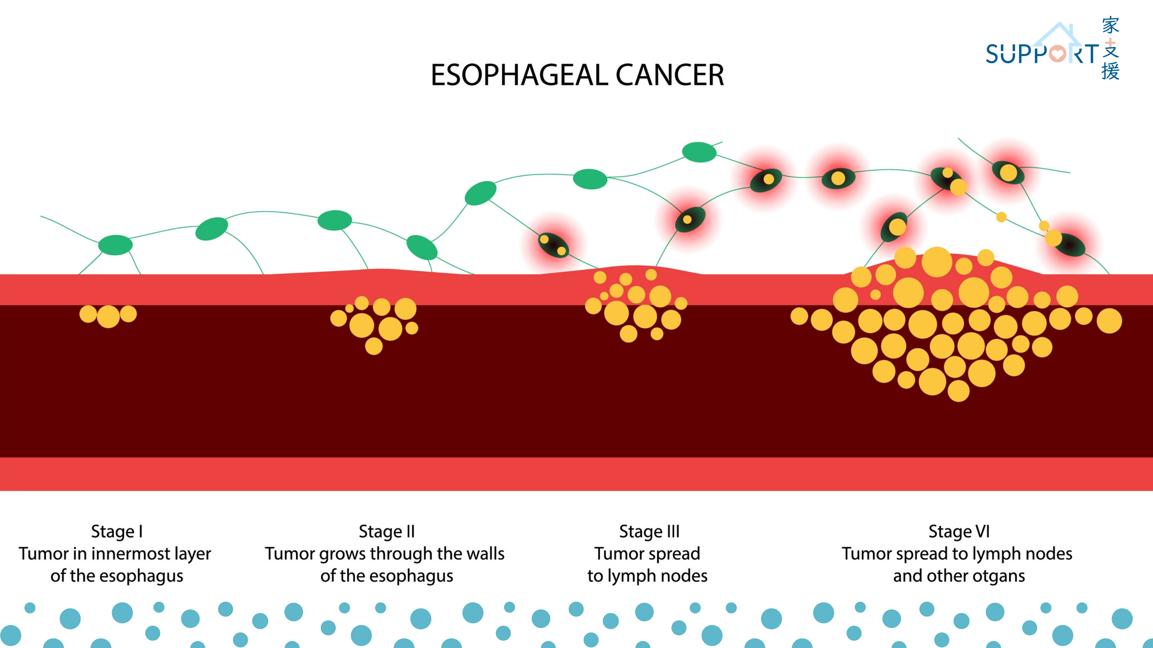
Stage I

The tumour has grown into the mucosa of the oesophagus.

Stage II

The tumour has grown into the muscle layer of the oesophagus, or has spread to 1 or 2 nearby lymph nodes.

Stage III

The tumour has grown outside the muscle layer of the oesophagus, or even beyond the wall of the oesophagus, or has spread to 3-6 nearby lymph nodes.

Stage IV

The cancer has spread to other parts of the body (distant metastasis), such as to the lungs, liver or stomach. This is also called metastatic oesophageal cancer.

Staging can be done by the following ways:

  • CT scan (Computed tomography)
    • A CT scan makes detailed cross-sectional images showing the tumour and the adjacent structures. It can also show if cancer has spread to organs near the pancreas, as well as to lymph nodes and distant organs.
    • Together with injection of intravenous contrast medium, the CT scan can find out exactly where the tumour is related to the nearby organs and blood vessels. This can help surgeon to evaluate whether the oesophageal cancer can be operable or not.
  • PET/PET-CT (Positron emission tomography)
    • PET-CT is a nuclear medicine imaging scan and uses radioactive isotopes to look for changes in the metabolic activity of body tissues. 
    • It is used to find out if the cancer has spread to other organs or tissues.

Treatment 

Treatment options and recommendations depend on several factors, including:

  • Type and stage of cancer 
  • Possible side effects 
  • The patient’s preferences 
  • Overall health

In cancer care, doctors of different specialties often work together to formulate an overall treatment plan that incorporates different types of treatments. This is called a multidisciplinary team.

Treatment for non-metastatic disease

Surgical treatment

Oesophagectomy

Oesophagectomy refers to the resection of the oesophagus, which is mainly performed for malignancy of the oesophagus. Occasionally, it is also indicated in benign conditions, like perforation and non-malignant narrowing (e.g. corrosive stricture). Following oesophagectomy, the stomach will be pulled up in order to regain the continuity of the gastro-intestinal tract. In certain cases, a segment of the large bowel is required to work as the conduit for reconstruction.

The operation is carried out under general anaesthesia with selective ventilation of the lungs. Epidural anaesthesia or patient-control-anaesthesia is frequently applied to reduce post-operative pain in view of the thoracotomy wound.

Conventionally, oesophagectomy includes three phases:

  • Surgical resection of the oesophagus
  • Mobilisation of the stomach keeping with it the blood supply
  • Anastomosis to maintain the continuity

Open surgical approach results in incisions over abdomen, the chest and neck. Nowadays, laparoscopic and thoracoscopic dissection can be performed as minimal invasive procedures. The surgery takes at least 5-6 hours to complete. After surgery, ICU care for ventilatory support and monitoring is the routine practice. Early ambulation and early oral feeding are advisable depending on the progress of recovery.  Post-operative intensive care is absolutely indicated. Specific complications related to oesophagectomy include:

  • Intra-operative bleeding in view of the extensive field of dissection and the nearby major vessels.
  • Anastomotic leakage because of tension to anastomosis and / or impaired blood supply.
  • Chylothorax as a result of damage to lymphatic system.
  • Chest infection or pneumonia. The majority of patients developing this complication are heavy smokers with poor respiratory function. The thoracotomy wound and single lung ventilation further hinder recovery. Indeed, sputum retention and chest complications are still the most likely causes of surgical failure.


Neoadjuvant/adjuvant treatment

Neoadjuvant treatment is an anti-cancer treatment given before definitive local treatment, such as surgery. For oesophageal tumours that are large or with nodal metastases, neoadjuvant treatment may be considered before the surgery to shrink the tumour (downstaging), thereby, increasing the possibility of a clean surgery (negative resection margins). Additional treatment may be considered after surgery (adjuvant treatment) for certain cases.

Neoadjuvant chemoradiation

Chemoradiation is commonly used for stage II to III oesophageal squamous cell carcinomas before surgery. The treatment lasts over a course of 5 weeks, at 5 days a week. Chemotherapy will be given during RT with weekly injection for 5 cycles, commonly used drugs include paclitaxel and carboplatin. The treatment can be delivered in an outpatient clinic without the need of hospital admission.

Figure: Radiotherapy planning for oesophageal cancer using CT scan

After completing the treatment, the disease will be re-evaluated by endoscopy and imaging to assess the response of the tumour. Surgery will be arranged within 1-2 months after completion of chemoradiation.

The resected tumour will be sent for examination under microscopy. If residual tumour cells are not found in the resected tumour (complete remission after chemoradiation), there will be no additional treatment required after surgery. For patients with residual disease noted in the resected cancer, recent clinical trial has shown that the use of immunotherapy after surgery (adjuvant nivolumab) will increase chances of survival. 

For some patients with residual disease left behind in surgery, usually due to the tumour being adjoint to important structures like the trachea or bronchus, additional radiation may be considered. Follow up treatment after surgery should be individualized depending on surgical and pathological findings, as well as the progress of recovery and health condition of the patient.


Neoadjuvant Chemotherapy

Squamous cell carcinomas of oesophagus

For squamous cell carcinoma that are long or with extensive nodal metastases, such as having spread to the neck or abdominal nodes, it may not be feasible to cover all sites noted on imaging in the radiotherapy field. For such patients, chemotherapy may be performed alone without radiation. The chemotherapy used will be more intensive, with combination of 2-3 drugs. Examples of combination chemotherapy include PF (Cisplatin-5FU) and TPF (cisplatin, 5 fluorouracil and docetaxel). After 2-3 cycles of chemotherapy, re-evaluation will be performed to assess its operability. 

Adenocarcinomas of gastroesphageal junction

Adenocarcinomas of gastroesophageal junction behaves more like stomach cancers and are managed similarly. These cancers are usually treated with peri-operative chemotherapy both before and after surgery. 

Chemotherapy will consist of 3 drugs (5-FU, oxaliplatin and docetaxel) (FLOT), used every 2 weeks for 4 cycles, followed by re-evaluation. After surgery, another 4 cycles of the same chemotherapy will be given.


Definitive chemoradiation

Chemoradiation may be used as a definitive treatment without surgery for squamous cell carcinoma, especially for patients who are medically unfit or unable to afford surgery.

For oesophageal cancers in the neck that are close to the larynx, definitive chemoradiation may be considered as alternative to surgery in order to preserve the larynx.

Definitive chemoradiation will involve high dose radiation for 5-6 weeks of daily treatment. Chemotherapy is commonly given during the course of radiation (2 cycles of cisplatin and 5-FU) and after completion of radiotherapy (another 2 cycles of cisplatin and 5-FU).

Treatment for advanced or metastatic disease

Systemic treatment

  • Chemotherapy

The mainstay of systemic treatment is palliative chemotherapy. Commonly used agents include cisplatin or oxaliplatin, 5 fluorouracil, docetaxel or paclitaxel. Single agent or combination of drugs can be considered.

  • Target therapy

For adenocarcinoma in the gastroesophageal junction, about 20-25% are HER2 positive. Trastuzumab is a monoclonal antibody against HER2 which can be used in combination with chemotherapy.

Another target therapy that can be considered for adenocarcinoma of the gastroesophageal junction is ramucirumab, which is inhibiting the formation of new blood vessels that supply nutrients for cancer growth. Ramucirumab can be used alone or in combination with chemotherapy.

  • Immunotherapy

For squamous cell carcinoma of oesophagus, anti-PD1 monoclonal antibodies may be used in combination with chemotherapy, or alone in tumours with expression of PD-L1.

For adenocarcinoma of gastroesophageal junction, anti-PD1 monoclonal antibodies can be used in combination with chemotherapy.

The combination or choice of drugs will be individualised depending on characteristics of the tumour, biomarker information, cost and toxicity of treatment, the patient’s health condition and preferences, etc. One’s choice of treatment should be discussed with a physician. 


Local treatment

Radiotherapy and oesophageal stent can be considered for relief of oesophageal obstruction. Painful bone metastases may be relieved by radiotherapy as well. 

Side effects

Side effects of chemotherapy include:

  • Decreased blood counts 
    • Blood monitoring is required during treatment. Some patients may need GCSF support during chemotherapy
  • Temporary hair loss 
    • Hair will grow post-chemotherapy
  • Immunosuppression and Infection
    • Patients with oesophageal cancer is particularly prone to chest infection. Patients should be aware of the risks of infection during chemotherapy and seek immediate medical attention if a fever or signs of infection appear. A prompt usage of antibiotics can be lifesaving. 

Side effects of radiotherapy include:

  • Skin reaction
    • irritated skin may be red and dry, which resembles a sunburn. In more severe cases, skin may start to peel. Skin reaction will subside in a few weeks after radiotherapy.
  • Pain while swallowing
    • This is due to reactions to radiation in oesophagus which will start to appear in the second week of radiotherapy, which will last until a few weeks after completion. Pain killers may be required. Ensuring sufficient nutrition intake during treatment is especially important for said patients, as apart from difficulty in swallowing due to obstruction caused by the tumour, pain during treatment worsen the situation. Tube feeding may be required in such cases.
  • Irritated lung and heart 
    • Radiation pneumonitis in the chest is a possible complication. Patients with poor lung function and extensive diseases are at higher risks.
  • Fistula formation
    • If the oesophageal tumour is close to or invading the trachea or bronchus, risk of fistula formation between the oesophagus and airway is enhanced, which will lead to repeated or persistent infection in the lung.

For patients treated with definitive chemoradiation, the irradiated oesophagus may be narrow due to fibrosis even if tumour is controlled. Such cases may be treated with endoscopic dilatation.

Prevention

The following may be effective in preventing oesophageal cancer:

  • Stopping tobacco use entirely
  • Avoiding alcoholic drinks with an alcohol content at above 20%
  • Reduce consumption of pickled or smoked food, including sauerkraut, preserved meats and betel nuts
  • Avoiding consumption of very hot food 
    • Do not consume until the food has slightly cooled
  • Treating existing symptoms of acid reflux
  • Increasing consumption of fresh fruits, vegetables and food with high fibre content
  • Maintaining a healthy body weight
    • Exercising for at least 30 minutes daily

Clinical trials in HKU

References

Smart patient (by Hospital Authority): Oesophageal cancer

American Society of Clinical Oncology (ASCO): oesophageal cancer

Canadian Cancer Society: oesophageal cancer


Special thanks to Mr. Joshua Tang, Ms. Sonia Yee-Sheung Chan (Class M23), medical student of Li Ka Shing Faculty of Medicine, the University of Hong Kong, and Prof. Dora Lai-Wan Kwong, Department of Clinical Oncology, the University of Hong Kong for authoring and editing this article.


Last updated on 18th Dec, 2023.