Skin Cancer

Skin Cancer
Background
Risk factors
Types
Symptoms
Diagnosis
Staging
Treatment
Treatment for recurrent or metastatic melanoma
Prevention
References
Share

Background

Skin cancer was common in Hong Kong, among which, non-melanoma skin cancer accounts for over 91.7%. Non-melanoma skin cancer ranked the ninth common among males in 2022. 

In 2022, there were 1,006 new cases of non-melanoma skin cancer, accounting for 2.84% of all new cancer cases in Hong Kong. The crude annual incidence rate of non-melanoma skin cancer per 100,000 Hong Kong population was 14.9. 

The incidence rate of skin cancer in Hong Kong is much lower than that of the Western countries. However, the local number of new non-melanoma skin cancer cases rose significantly by 10.8% from 2012 to 2022. 

Although the incidence of skin cancer is high, its mortality rate is relatively low. Since the skin is the most visible organ, most abnormalities can be easily detected. There were total 29 people died from non-melanoma skin cancer in 2022, accounting for 0.2% of all cancer deaths.

If any unusual skin changes are noticed, please visit a doctor as soon as possible. Early detection increases the chance of successful treatment.


What is skin cancer?

  • Structure and functions of the skin
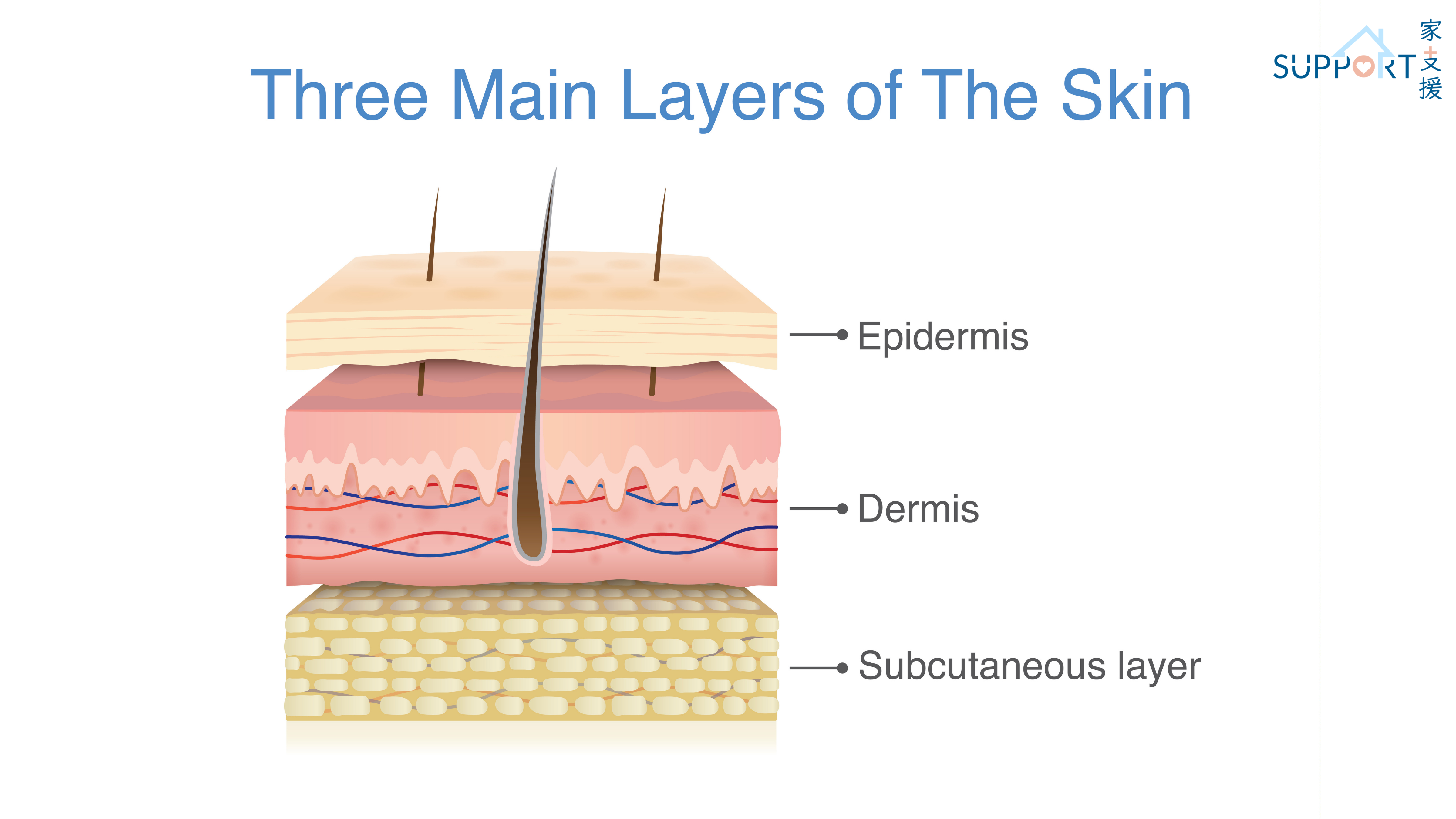
    • The skin is the largest organ of the body. It consists of three parts: epidermis, dermis and subcutaneous tissue.
    • The epidermis consists of three types of cells: keratinocytes, Langerhans cells, and melanocytes.
    • The skin protects the body from foreign agents, lowers the temperature by sweating and prevents dehydration.
  • Carcinogenesis
    • Skin cancer is mainly caused by ultraviolet rays (UV).
    • The main source of ultraviolet rays is sunlight. The second is UV lamps from fitness and beauty centres.
    • There are three wavelengths of UV, UVA, UVB and UVC. While UVB is the main mutagenic cause of basal cell carcinoma and squamous cell carcinoma, UVA, which is released in large amounts by tanning beds, mainly causes melanoma.
    • UV-related damage to skin is accumulative. Even without sunburn, skin tissues will still be injured after prolonged exposure.
    • Previous studies suggested that if skin cells have been damaged at a young age, lack of proper UV protection in adulthood will cause the damaged cells to mutate.
    • Skin cancer often appears in areas that are exposed to sunlight, such as the face, lips and neck. However, it can also appear in areas not likely to be exposed to sunlight, such as the palms, soles and gaps between toes.
    • Skin cancer which occurs in areas away from sunlight may have genetic factors.

Risk factors

  • Number of moles, which is more than usual
  • Presence of macule
  • Family history of melanoma
  • Long working hours under the sun
  • Frequent sunburns, especially with peeling skin
  • Frequent use of tanning beds
  • Light-coloured skin that is hard to tan but can easily be sunburnt
  • Red or blonde hair with light-coloured pupils
  • Immunocompromised e.g., received organ transplantation or HIV positive
  • Burnt, injured skin or receiving treatment for other skin diseases
  • Close contact with chemical carcinogens

Types

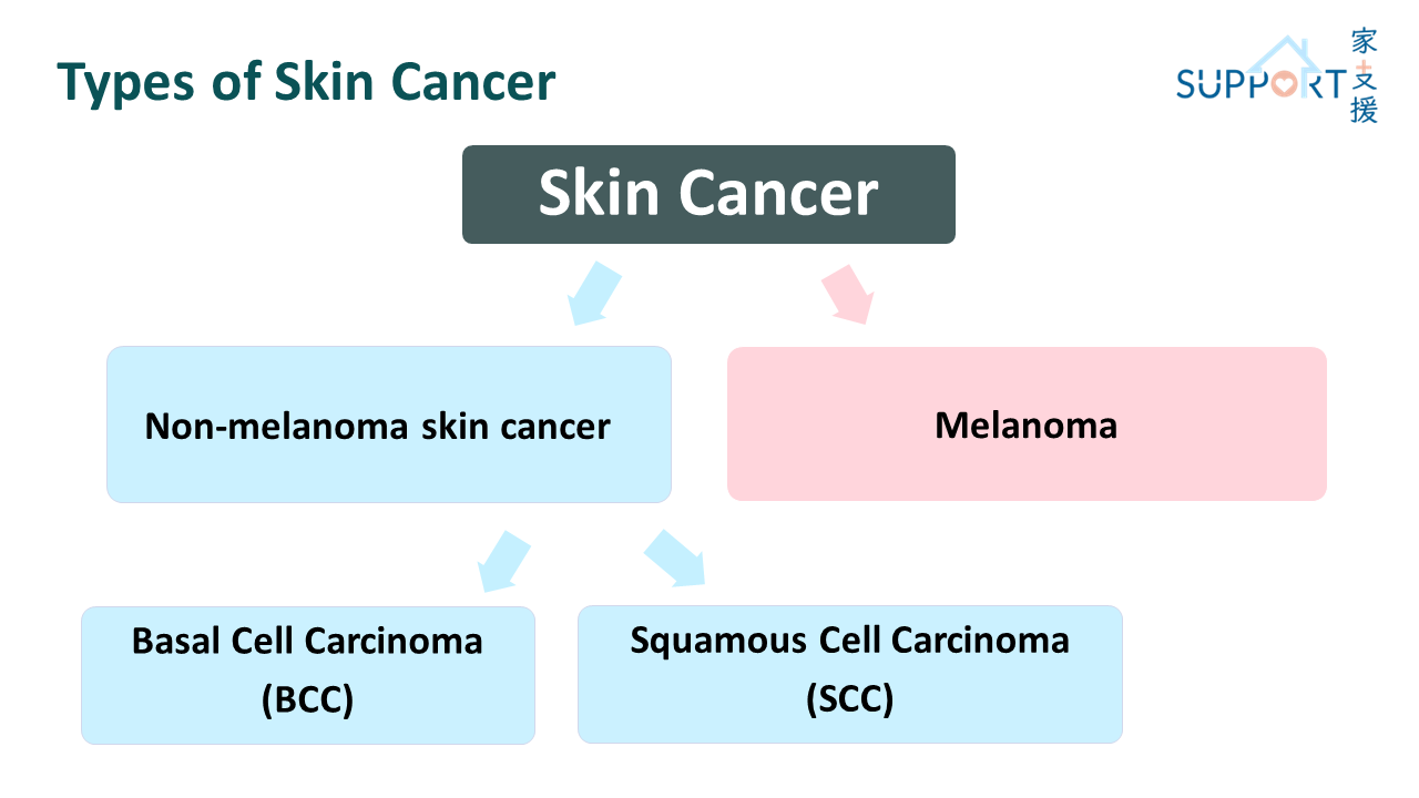
Skin cancers are classified into:

  • Non-melanoma skin cancer
    • Basal cell carcinoma
    • Squamous cell carcinoma
  • Melanoma


Non-melanoma skin cancer

  • Non-melanoma skin cancer includes basal cell carcinoma and squamous cell carcinoma.
  • Basal cell carcinoma and squamous cell carcinoma make up 98% of skin cancers.
  • Non-melanoma skin cancer is the most common malignant tumour seen in Caucasians. Approximately one million cases were diagnosed in the US in 1999. One out of five Americans will be diagnosed with skin cancer in their lifetime.

Melanoma

  • Melanoma is a highly malignant tumour caused by the mutation of melanocytes in the epidermis.
  • It is highly lethal and often metastasises early.
  • Like non-melanoma skin cancer, the number of melanoma cases worldwide is rapidly increasing. It is more commonly seen in Caucasians than in Black people.

Symptoms

Characteristics of basal cell carcinoma

  • Often appears in head and neck regions which are exposed to sunlight (60%-80%) and the nose (30%).
  • Pain, bleeding and blood clot(s) are often observed at the pathological site which do not heal on their own. Itchiness, squamous-like flat scaly clots and painless ulceration are also common.
  • Basal cell carcinoma is usually seen as carcinoma-in-situ. It grows along organs with weaker immune defenses. It is slow in growing and seldom metastasises, hence curable if discovered early.
  • Carcinoma-in-situ can cause infection, necrosis, pain and irreversible damage to local structures.


Characteristics of squamous cell carcinoma

  • 30% of all skin cancers are squamous cell carcinomas.
  • Usually arises from skin exposed to strong UV light, skin with previous burn injury, or tissue with slow recovery.
  • Typically originates in elderly at sites exposed under sunlight, such as the face, hands and forearms. Most cases are accompanied with rashes, masses and ulceration.
  • Squamous cell carcinoma grows slowly, hence it is usually curable. However, it has to be managed carefully to prevent metastasis.
  • Squamous cell carcinoma is more aggressive than basal cell carcinoma. It usually penetrates the basal lamina. It commonly metastasises through the lymphatic system to the lymph nodes and less commonly spreads to distant organs like lungs or bones.


Characteristics of melanoma

  • Melanoma accounts for around 2% of all skin cancers.
  • Commonly presents as a rapidly forming deposit of melanin with ulceration, but can sometimes be amelanotic.
  • In Caucasian ladies, 50% of melanomas occur in the legs; in Caucasian gentlemen, most melanomas are found on the trunk. 
  • In Asians, melanomas mostly arise in the soles, palms, nail beds or mucosa. 
  • Chance of melanoma in the face increases with age. In middle age, it is more commonly observed in other parts of the body. 
  • Melanoma can easily spread. Common metastatic sites include local lymph nodes, lungs and brain. Therefore, it is important to diagnose melanomas as early as possible.
  • Symptoms of melanoma:
    • New, unusual looking pigmented area on skin 
    • Itchiness, pain and inflammation of the pathogenic site
    • Prolonged unhealed wounds 
    • Growth, transformation or colour change in moles or pigmented area
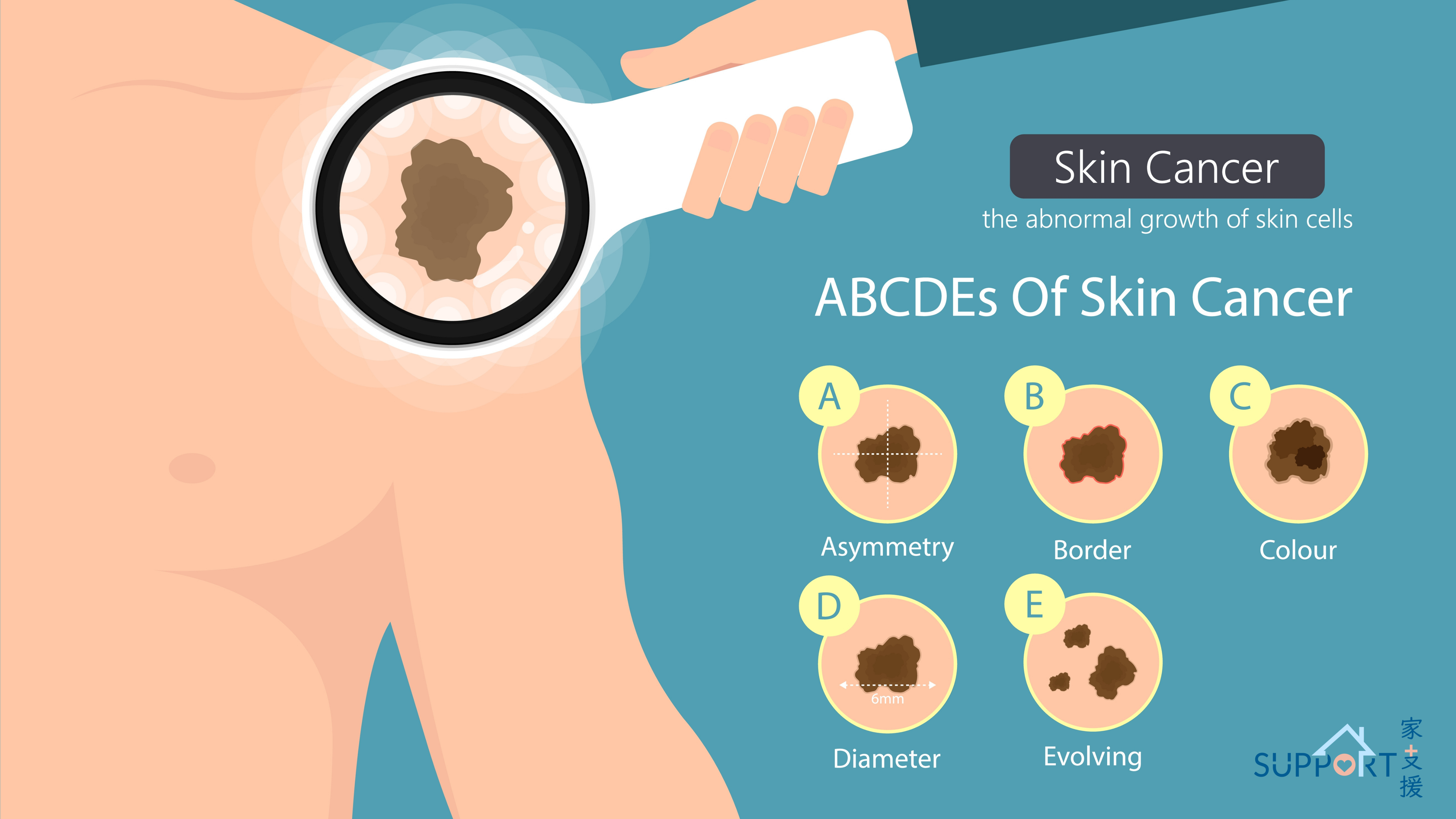
    • Transformation of moles:
      • Asymmetric shape 
      • Irregular borders
      • Colour change
      • Size above 6mm in diameter or observable growth
      • Asymmetric bulging

Diagnosis

Biopsy is required to diagnose different types of skin cancer. The doctor will cut out a small area of the suspected site under local anaesthesia. The cells will be sent to the laboratory to identify if the tumour is benign or malignant. The process will take around two weeks.

After getting the diagnosis, doctor will check the exact location, size and degree of metastasis of the skin cancer. Since skin cancer usually grows on the skin surface and the pace of growth is slow with a low chance of metastasis, further investigations may not be required.

The doctor will palpate for any swelling or enlargement of the lymph nodes near the skin cancer. If necessary, further investigations, e.g. blood tests, X-rays, or computed tomography (CT) may be performed.

If malignant melanoma is confirmed, you may need to have more detailed investigations, e.g. CT scan to check for any distant spread.

Staging

Staging for melanoma 

  • Stage 0
    • This refers to melanoma in situ, which means melanoma cells are found only in the outer layer of skin or epidermis. 
    • This stage of melanoma is very unlikely to metastasise to other parts of the body.
  • Stage I
    • Stage I melanoma only grows on the skin and is relatively thin. 
    • Stage I is further divided into 2 subgroups, IA or IB, according to the thickness of the melanoma and whether ulceration is seen under a microscope.
  • Stage II
    • Stage II melanoma is thicker than stage I melanoma, extending into the epidermis, the dermis and subcutaneous tissues. 
    • It has a higher chance of spreading. 
    • Stage II is further divided into 3 subgroups, A, B, or C, according to the thickness of the melanoma and whether ulceration is seen.
  • Stage III
    • Stage III melanoma is one that has metastasised locally, through the lymphatic system to a regional lymph node (near where the cancer started), or to a skin site on the way to a lymph node.
    • Stage III is further divided into 4 subgroups, A, B, C, or D, according to the size and number of lymph nodes involved with melanoma, whether the primary tumour has satellite, microsatellite and/or in-transit lesions, and if it appears ulcerated under a microscope.
  • Stage IV
    • Stage IV melanoma has metastasised through the bloodstream to other parts of the body, such as distant locations on the skin or soft tissue, distant lymph nodes, or other organs like the lung, liver, brain, bone, or gastrointestinal tract. 
    • Stage IV melanoma is further evaluated based on the location of distant metastasis:
      • M1a: The cancer has only spread to distant skin and/or soft-tissue sites.
      • M1b: The cancer has spread to the lung.
      • M1c: The cancer has spread to any other location that does not involve the central nervous system.
      • M1d: The cancer has spread to the central nervous system, including the brain, spinal cord, and/or cerebrospinal fluid, or lining of the brain and/or spinal cord.
  • Recurrent
    • Recurrent melanoma is a type of melanoma that comes back after treatment. 
    • The doctor will conduct detailed tests and scans to identify the extent of the recurrence. 

Treatment

Treatment for skin cancers is mainly by surgery and radiotherapy.

Treatment for basal cell carcinoma

  • Majority of basal cell carcinoma can be cured with surgery with or without radiotherapy.
  • Radiotherapy may be given in the following conditions:
    • Patients who are not able or do not want to have surgery o
    • Tumours on the eyelids, nose, or ears, which can be hard to treat surgically
    • Some older patients if curing the cancer may not be as important as controlling it over the long term and limiting side effects
    • After surgery if there is a risk that not all the cancer cells have been removed, or if there is a high risk that the cancer might come back 
    • Occasionally used when the cancer has spread to other places, such as lymph nodes or the lungs
    • However, radiotherapy usually cannot be repeated in the same area if it was given during primary treatment due to higher risk of serious side effects
  • The doctor may also recommend other treatment options such as cryotherapy, photodynamic therapy and topical medications (e.g. imiquimod, 5-fluorouracil) according to the patient's condition.
  • Patients should be followed up in the long run as there is a 5-10% chance of recurrence and metastasis.
  • In case of advanced basal cell skin cancer with metastases to nearby or distant lymph nodes and/or organs, in addition to surgery and radiotherapy, systemic therapy such as immunotherapy (cemiplimab) or targeted therapy (vismodegib) may be required sequentially or as alternative. 


Treatment for squamous cell carcinoma

  • Majority of squamous cell carcinoma can be found and treated at an early stage with local treatments such as surgery and radiotherapy. Small squamous cell carcinomas can usually be cured with these treatments. 
  • Topical medications used in basal cell carcinoma can also be used for squamous cell carcinoma, with additional options such as calcipotriene (a form of vitamin D), urea, lactic acid, or salicylic acid (keratolytics), diclofenac (anti-inflammatory), trichloroacetic acid, acitretin and isotretinoin (retinoids).
  • While it is not common, squamous cell carcinoma can sometimes spread to lymph nodes or distant parts of the body. If this happens, surgical removal of nearby lymph nodes may be performed. Moreover, systemic therapies such as targeted therapy (EGFR inhibitor cetuximab), immunotherapy (cemiplimab, pembrolizumab, nivolumab) with out without concurrentchemotherapy (platinum, paclitaxel, 5-fluorouracil, capecitabine) may be given as systemic treatment.
  • Patients are required to be followed up in the long run as there are chances of recurrence and metastasis.


Treatment for melanoma

Treatment for localised melanoma 

  • Surgery
    • Surgery is the main treatment for localised melanoma.
    • Patients will require a skin graft or skin flap when the melanoma is too thick.
    • Some patients need to have their lymph nodes in the region near the primary melanoma tumour removed to prevent recurrence or metastasis.
    • Patients should be followed up in the long run as there are chances of recurrence and metastasis.
  • Radiotherapy
    • External beam radiation therapy (EBRT) may be an option to treat some early-stage melanomas, if surgery cannot be done for some reason. 
    • Radiotherapy may be used after surgery as adjuvant treatment if there is a high risk local recurrence. Sometimes, adjuvant radiotherapy may be given in the area where lymph nodes were removed, especially if many of the nodes contained cancer cells. 
    • Side effects of EBRT: sunburn-like skin problems, changes in skin colour, hair loss in the area where radiotherapy enters the body, fatigue, nausea if aimed at the abdomen
  • Immunotherapy
    • PD-1 (Programmed Death-1) inhibitors and CTLA-4 (Cytotoxic T Lymphocyte Associated protein-4) inhibitor are immune checkpoint inhibitors which are used in early-stage melanoma before and/or after surgery.
    • Pembrolizumab (PD-1 inhibitor), nivolumab (PD-1 inhibitor), ipilimumab (CTLA-4 inhibitor) enable white blood cells to recognise and attack cancer cells thereby decreases the chance of recurrence and increases survival rates of melanoma patients.
    • Neoadjuvant (before surgery) regimens: 
      • Pembrolizumab (Keytruda)
      • Nivolumab (Opdivo) and ipilimumab (Yervoy) combination 
      • Nivolumab (Opdivo) 
      • Nivolumab and relatlimab combination (Opdualag)
    • Adjuvant (after surgery) regimens:
      • Nivolumab (Opdivo)
      • Pembrolizumab (Keytruda)
    • Administration: via intravenous route once every 2 to 6 weeks, for one year 
    • Side effects: fatigue, cough, nausea, skin rash, poor appetite, constipation, joint pain, diarrhoea
    • Rare but more serious side effects: infusion reactions (fever, chills, flushing of the face, rash, itchy skin, feeling dizzy, wheezing, and trouble breathing), autoimmune reactions (serious or even life-threatening problems in the lungs, intestines, liver, hormone-making glands, kidneys, or other organs)
  • Targeted therapy
    • Use of the combination of a BRAF inhibitor and MEK inhibitor i.e. dabrafenib (Tafinlar) and trametinib (Mekinist) after surgery decreases the chance of recurrence and increases survival rates in patients with BRAF V600 mutation. 
    • Administration: tablets or capsules, taken orally, once or twice daily, for total 12 months

Treatment for recurrent or metastatic melanoma

  • Most recurrence will spread to other parts of the body. Therefore, the survival rate is low.


Chemotherapy

  • Chemotherapy may be considered for metastatic melanoma patients as subsequent treatment after first-line therapy with newer forms of immunotherapy and targeted drugs which are typically more effective.
  • Examples of chemotherapy agents: dacarbazine, temozolomide, paclitaxel, carboplatin
  • However, chemotherapy only poses a moderate effectiveness while posing chances of a number of serious side effects, such as hair loss, mouth sores, loss of appetite, nausea and vomiting, diarrhoea or constipation, increased risk of infection, easy bruising or bleeding, fatigue, peripheral neuropathy.
  • Administration: via intravenous or oral route. 


Targeted therapy

1. BRAF inhibitors E.g., Vemurafenib (Zelboraf), Dabrafenib (Tafinlar), and encorafenib (Braftovi)

  • 25% of patients with melanoma carry BRAF mutations in Asia. These patients can be treated by BRAF inhibitors Vemurafenib (Zelboraf), Dabrafenib (Tafinlar) and encorafenib (Braftovi) which reduce the size of the tumour, slow its growth, and improve survival. It is essential to have genetic test confirming the presence of BRAF V600 mutation before using this group of medications.
  • Administration: tablets or capsules, taken orally, once or twice a day.
  • Common side effects: Skin thickening, rashes, itchiness, photosensitivity, headache, fever, joint pain, fatigue, hair loss and nausea.
  • Less common but serious side effects: arrhythmias, liver disease, kidney failure, severe allergic reactions, severe skin problems, eye problems and increase in blood sugar levels. Some patients may develop squamous cell carcinoma after taking the drug, but they are usually milder than melanoma and can be removed surgically.


2. MEK inhibitors E.g., Trametinib (Mekinist)

  • MEK gene works together with BRAF gene to stimulate cancer cells to proliferate. With this mechanism, MEK inhibitor (e.g. Trametinib) is useful in inhibiting tumour growth in melanoma patients with BRAF V600E mutations.
  • Administration: tablets or capsules, taken orally, once or twice a day.
  • Common side effects: Rashes, nausea, diarrhoea, edema and photosensitivity.
  • Less common but serious side effects: Heart damage, massive bleeding, blindness, lung problems and skin infections.
  • The combination of MEK inhibitors and BRAF inhibitors are commonly used for patients with metastatic melanoma with BRAF V600 mutation. The combination regimen, compared to a single agent, is more effective and has fewer side effects (such as developing other skin cancers).
    • Dabrafenib (Tafinlar) and trametinib (Mekinist)
    • Encorafenib (Braftovi) and binimetinib (Mektovi)


3. Other targeted therapies

  • A small proportion of metastatic melanoma patients express the following gene mutations which can be treated by the corresponding targeted therapies below: 

Mutation 

Targeted therapy drug 

BRAF fusions and non-V600 mutations 

Trametinib (Mekinist) 

C-KIT gene mutation 

  • Imatinib (Gleevec or Glivec)
  • Nilotinib (Tasigna)
  • Dasatinib (Sprycel)
  • Ripretinib (Qinlock) 

NRAS mutation 

Binimetinib (Mektovi) 

ALK fusion 


ROS1 fusion 

  • Crizotinib (Xalkori)
  • Entrectinib (Rozyltrek) 

NTRK fusion 

  • Larotrectinib (Vitrakvi)
  • Entrectinib (Rozyltrek) 


Immunotherapy

  • PD-1 inhibitors, PD-L1 (Programmed Death Ligand-1) inhibitor, and CTLA-4 inhibitor are immune checkpoint inhibitors which are effective in treating end stage melanoma.
  • Pembrolizumab (PD-1 inhibitor), nivolumab (PD-1 inhibitor), atezolizumab (PDL1 inhibitor), and ipilimumab (CTLA-4 inhibitor) enable white blood cells to recognise and attack cancer cells thereby prolonging the lifespan of melanoma patients.
  • First-line regimens:
    • Nivolumab (Opdivo) and ipilimumab (Yervoy) combination
    • Pembrolizumab (Keytruda) ±  ipilimumab (Yervoy)
    • Nivolumab (Opdivo)
  • Subsequent-line regimens:
    • Ipilimumab (Yervoy)
    • Pembrolizumab (Keytruda) and lenvatinib (Lenvima, multi-kinase inhibitor) combination
  • Administration: via intravenous route, once per two to three weeks.
  • Side effects: Immune checkpoint inhibitors have fewer side effects when compared with older generation drugs. The common side effects are fatigue, coughing, vomiting, rashes, itchiness, constipation, loss of appetite, joint pain and diarrhoea.
  • The immune checkpoint inhibitors reactivate the body's self-defense mechanisms to fight cancer cells. However, in rare cases, these drugs might prompt the immune system to attack other normal organs and tissues in the body, e.g., lung, intestine, liver, kidneys and bowel, leading to serious and even fatal side effects.


Radiotherapy

Palliative radiotherapy can be used to relieve pain and control bleeding in patients with advanced melanoma. It is not expected to cure the cancer, but it may help shrink it or slow its growth for a time to help control some of the symptoms. 

Prevention

  • Studies have shown that 80% of skin cancers can be prevented by protective measures against sunlight.
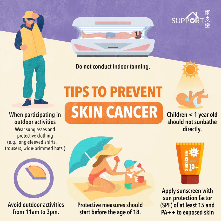
  • Protective measures should start early as 80% of damage by ultraviolet rays is done before the age of 18.

  • Children under one year old should not sunbathe directly.
  • Do not conduct indoor tanning.
  • Avoid outdoor activities from 11am to 3pm as ultraviolet rays are the most damaging during this period. Persons with pale complexions might get sunburnt in less than 15 minutes at noon.
  • Wear protective clothing such as long-sleeved shirts, trousers and wide-brimmed hats when participating in outdoor activities. Wear sunglasses when necessary.
  • Apply sunscreen with sun protection factor (SPF) of at least 15 and PA++ to exposed skin when participating in outdoor activities to protect the skin from UVA and UVB.
    • SPF refers to the ability of a sunscreen to protect the skin from UVB. The higher the number, the better the protection.
    • SPF = Time required for skin to turn red from UVB after applying sunscreen/Time required for skin to turn red from UVB without applying sunscreen.
    • Sunscreen with SPF15 can block 93% of UVB, while sunscreen with SPF30 can block 97% of UVB.
    • PA refers to the ability of a sunscreen to protect our skin from UVA. The greater number of ‘+’, the better the protection.
    • Tips for applying sunscreen
      • One teaspoon of sunscreen is required for the face while 30mL of sunscreen is required for the whole body.
      • Use sufficient sunscreen with SPF 15/30, rather than a thin layer of sunscreen with SPF 60/100.
      • Apply sunscreen 30 minutes before outdoor activities.
      • Apply sunscreen when swimming outdoors.
      • Reapply sunscreen every 2 hours when staying outdoors for a long period of time.
      • Reapply sunscreen after swimming or sweating.
      • Seek shade whenever possible even with sunscreen applied.

References

American Society: Skin Cancer

Cancer Council: Skin Cancer

Macmillan Cancer Support: Skin cancer

NCCN Guidelines for Patients. Basal Cell Skin Cancer, 2022.  

NCCN Guidelines for Patients. Melanoma, 2024.  

NCCN Guidelines for Patients. Squamous Cell Skin Cancer, 2023.  

NCCN Guidelines. Basal Cell Skin Cancer. Version 1.2025. 

NCCN Guidelines. Melanoma: Cutaneous. Version 1.2025. 

NCCN Guidelines. Melanoma: Uveal. Version 1.2025. 

NCCN Guidelines. Squamous Cell Skin Cancer. Version 1.2025. 

Smart Patient (by Hospital Authority): Skin cancer 


Special thanks to Ms. Locani Hing-Wai Wong (Class M25), Mr. Alex Yung-Pok Lee (Class M25), medical student of Li Ka Shing Faculty of Medicine, the University of Hong Kong, and Dr. Wendy Wing-Lok Chan, Department of Clinical Oncology, the University of Hong Kong for authoring and editing this article.


Last updated on 19th Jan, 2025.