Malignant Pleural Effusion

Malignant Pleural Effusion
What is it?
Causes
Symptoms
Diagnosis
Treatment
References
Share

What is it?

How is it different from pleural effusion?

Malignant pleural effusion is the buildup of fluid that collects between the lung and the chest wall caused by the spreading of cancer cells.

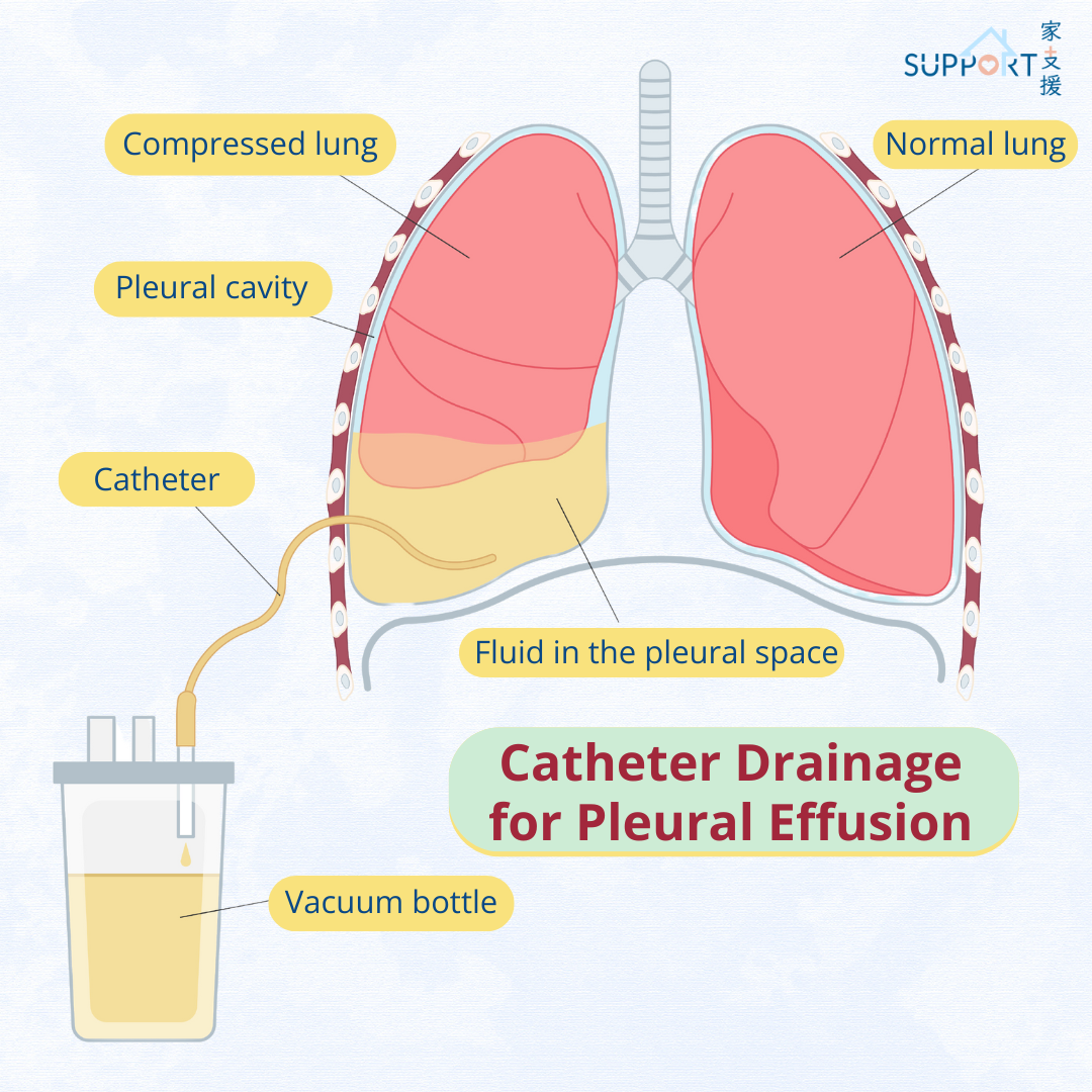
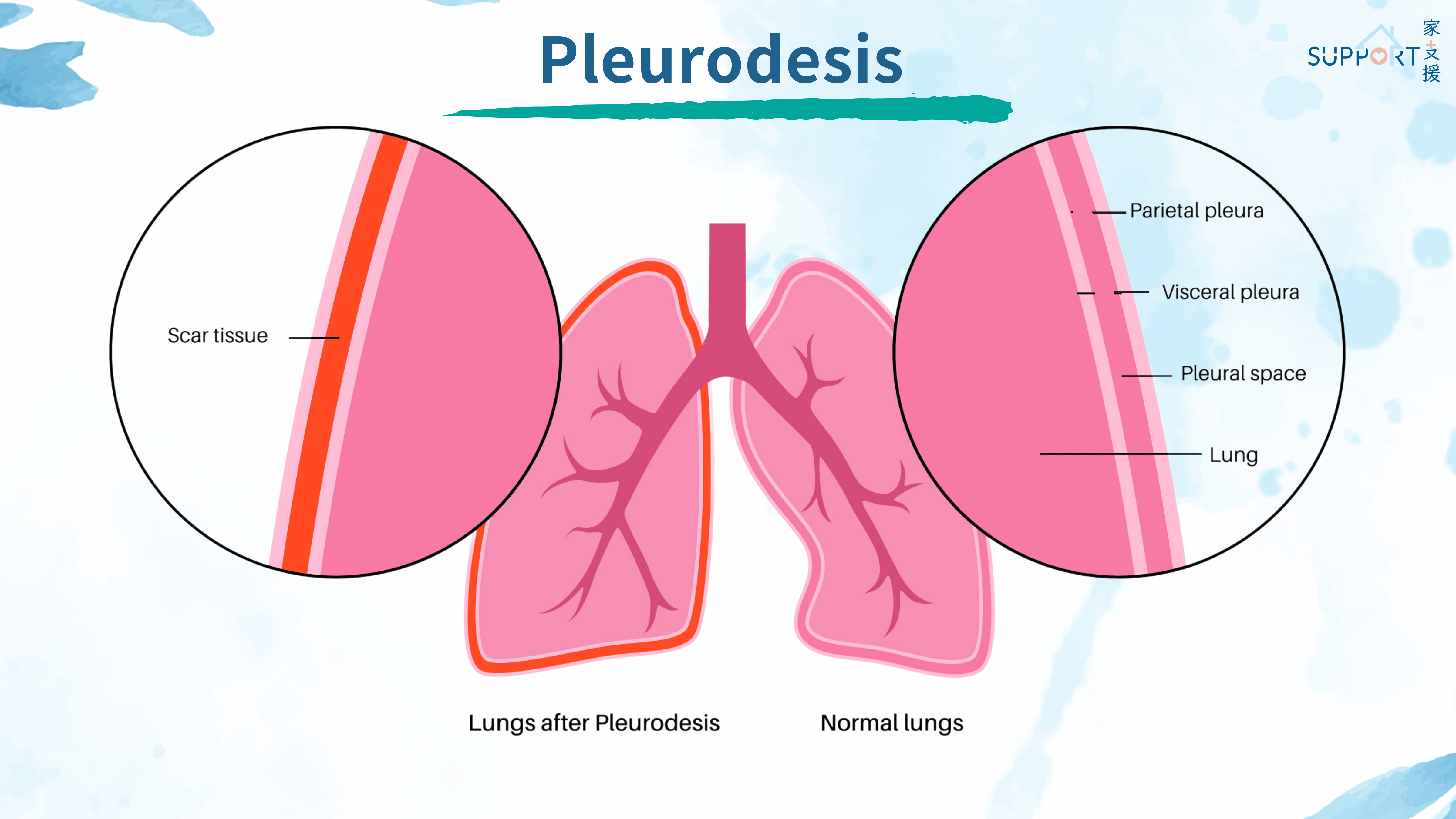
There are two layers of tissues that cover and protect the lungs. They are called pleura (or pleural membranes). Between the 2 pleura is a space called the pleural space, which contains a very small amount of fluid (approximately 1 teaspoon) that helps the lungs move easily during breathing. Pleural effusion occurs when there is excessive fluid buildup in the pleural space. This makes the expansion of lungs more difficult, leading to difficulty in breathing.

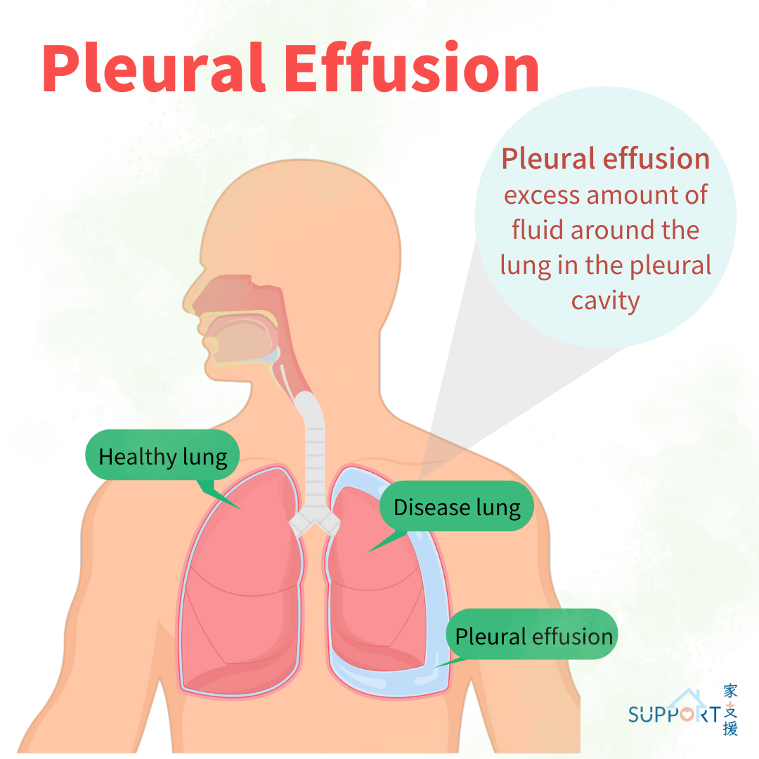
Malignant pleural effusion is a specific type of pleural effusion caused by cancer. The cancer cells increase the production of pleural fluid and decrease the absorption of the fluid, causing an accumulation of fluid.


Causes

What cancer types can cause malignant pleural effusion?

Any cancer types that spread to the pleura can cause malignant pleural effusion. Certain types of cancer are more likely to cause malignant pleural effusion. These include:

  • Lung cancer
  • Mesothelioma (cancer of the pleura)
  • Ovarian cancer
  • Breast cancer
  • Colorectal cancer
  • Lymphoma

Symptoms

Common symptoms of malignant pleural effusion include:

  • Shortness of breath/ breathlessness: the most common and usually the first symptom
  • Cough
  • Chest pain, especially during deep breathing
  • Fatigue

Diagnosis

How is a pleural effusion diagnosed?

Investigations for suspected malignant pleural effusion usually include two parts:

  1. Confirming the diagnosis of pleural effusion
  2. Working up on the underlying cause of malignant pleural effusion.

To diagnose pleural effusion, your doctor will peform:

  • Chest X ray: A film showing the view of a chest, including heart and lungs.
  • Thoracic ultrasound (TUS): Provides radiation-free images of the pleural space, also helps guiding thoracentesis and pleural biopsy.
  • Chest computed tomography (CT) scan: Providing more detailed information on the collection of fluid, including the adjacent structures in the chest and identifying source of primary cancer.

To find out the cause of malignant pleural effusion, your doctor will need to take a sample of fluid to check for the cause. This can be done by the following ways:

Pleural fluid aspiration (Thoracentesis)

  • This is a simple procedure done by inserting a small needle or tube into the pleural space to remove the fluid accumulated there. The fluid aspirated out will be tested to determine the cause of effusion and look for cancer cells.
  • The procedure is performed while you are sitting up and leaning forward on a table. The skin is disinfected and your doctor will use a fine needle to give you some local anaesthesia to reduce pain and discomfort. Then your doctor will make a small cut in the skin and insert the plastic tube to the pleural space under ultrasound guidance. The fluid will then be drained out.
  • Risk: The procedure is safe but there may still be risk of air in sucking into the lung (pneumothorax), bleeding, infection, etc.

Pleural biopsy

  • This is a more invasive procedure that takes a piece of pleura for detailed analysis. It can be done by needle biopsy, thoracoscopic biopsy or open biopsy.
  • For thoracoscopic biopsy, you will have the procedure under general anaesthesia. Your doctor will put a flexible, lighted tube mounted with a camera into the pleural space. Biopsy will be taken and sent for analysis under a microscopy.

Treatment

How to manage malignant pleural effusion?

The doctor will formulate a management plan for malignant pleural effusion based on the severity of the effusion.

  • Mild/ asymptomatic malignant pleural effusion: Patients can be managed by observation alone.
  • Moderate to severe malignant pleural effusion: The goal of management is to alleviate the shortness of breath by removing the excessive fluid by pleural drainage and indwelling pleural catheter (IPC), and performing treatment to stop fluid building up.

1. Pleural drainage

  • Your doctor will use a thoracic ultrasound scan to identify the best location to insert the chest drain tube.
  • You will be asked to sit on the edge of the bed or on a chair and lean forward over a table with a pillow on it for better exposure of your back.
  • Your doctor will then clean the skin over the area (usually in the side of the chest) with antiseptic solution and inject a small amount of local anaesthetic, minimising the pain throughout the procedure.
  • Then your doctor will make a small cut on your skin and insert a thin catheter into the pleural space under ultrasound guidance. After confirming the catheter’s position, your doctor will connect the catheter with a collecting bag or bottle which is kept lower than your chest and fluid drains out automatically.
  • The catheter will be sutured and secured in place.
  • Since the maximum amount of fluid drained per day is around 1-1.5L, it may take a few days to drain all the accumulated fluid from the pleural space.
  • Once all the fluid is drained, your doctor or nurse will take the catheter out.
  • Potential complications of pleural drainage include bleeding, blockage of drain, infection, direct injury to the lungs and abnormal air collecting in pleural space (pneumothorax).


2. Indwelling pleural catheter (IPC)

  • If the pleural fluid keeps coming back, some patients may have the indwelling pleural catheter (IPC) insertion to get back home for repeated drainage and avoid repeated chest tube insertion.
  • It has a long silicon tube that is inserted into the pleural cavity and has a value on the end preventing fluid leaking out of the tube and air from leaking in.
  • IPC is indicated for patients with recurrent malignant pleural effusion and those who are unable have repeated visits to hospitals for pleural fluid management.
  • Possible complications of IPC include infection at the insertion site (cellulitis) or within the pleural space (empyema), blockage and displacement of catheter, pleural inflammation, pneumothorax and cancer cells spreading along the catheter tract (catheter tract metastasis).


3. Pleurodesis

  • Pleurodesis is a procedure that seals the pleural space to stop fluid from building up. It is commonly done after drainage of the pleural fluid.
  • There are two types of pleurodesis: Chemical pleurodesis and surgical pleurodesis.
  • Chemical pleurodesis is done by injecting sterile powder into the pleural space through the drainage catheter or chest drain. The doctor will them clamp the tube. The solution will cause the pleura sticking together.
  • Surgical pleurodesis is done by a thoracoscopy. The surgeon brushes the pleural membranes to cause abrasions, scarring and inflammation which help to adhere the layers of pleura together.

4. Managing the primary cancer

  • Your doctor will formulate a management plan on the primary cancer causing malignant pleural effusion. Management options will depend on the type and stage of the primary cancer, including chemotherapy, radiotherapy, immunotherapy, and/or targeted therapy.